Fraud Blocker Skip to main content

Proteins Collection (page 9)

Proteins: The Building Blocks of Life From the intricate network of nerve and glial cells to the mesmerizing patterns seen under a light micrograph

Background imageProteins Collection: Amyloid precursor protein molecule

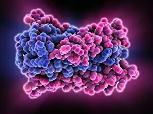
Amyloid precursor protein molecule
Amyloid precursor protein. Molecular model showing the structure of the protease inhibitor domain of an amyloid precursor protein (APP)
Background imageProteins Collection: Amyloid precursor protein molecule C014 / 0863

Amyloid precursor protein molecule C014 / 0863
Amyloid precursor protein. Molecular model showing the structure of the protease inhibitor domain of an amyloid precursor protein (APP)

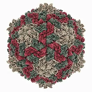
Background imageProteins Collection: Ryegrass mottle virus capsid

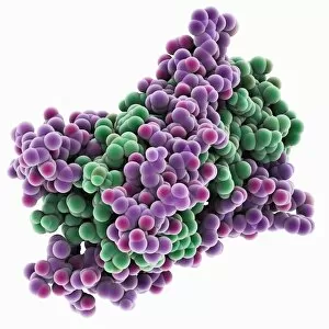
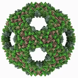
Ryegrass mottle virus capsid, molecular model. This plant virus is named for its infection of ryegrass, and the discolouration it causes

Background imageProteins Collection: Norwalk virus capsid, molecular model

Norwalk virus capsid, molecular model. This norovirus, which causes a viral form of gastroenteritis, is transmitted from person-to-person or through contaminated food

Background imageProteins Collection: Semliki forest virus capsid

Semliki forest virus capsid, molecular model. This virus, named for the forest in Uganda where it was identified, is spread by the bite of mosquitoes. It can infect both humans and animals

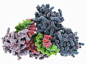
Background imageProteins Collection: TATA box-binding protein complex C014 / 0879

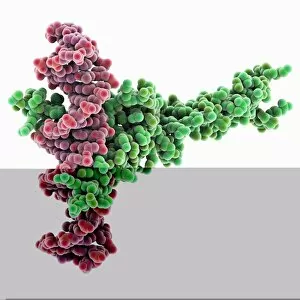
TATA box-binding protein complex C014 / 0879
TATA box-binding protein complex. Molecular model showing a TATA box-binding protein (TBP, lilac) complexed with a strand of DNA (deoxyribonucleic acid, green and red)

Background imageProteins Collection: Physalis mottle virus capsid

Physalis mottle virus capsid
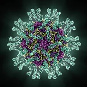
Avian polyomavirus capsid, molecular model. This virus, one of a range named for their potential to cause multiple tumours, infects birds. Discovered in budgerigars in 1981, it is often fatal

Background imageProteins Collection: Methyladenine glycosylase bound to DNA C014 / 0877

Methyladenine glycosylase bound to DNA C014 / 0877
Methyladenine glycosylase bound to DNA. Computer model showing a molecule of human DNA-3-methyladenine glycosylase (purple) in complex with DNA (deoxyribonucleic acid, green and orange)

Background imageProteins Collection: Type I topoisomerase bound to DNA C014 / 0883

Type I topoisomerase bound to DNA C014 / 0883
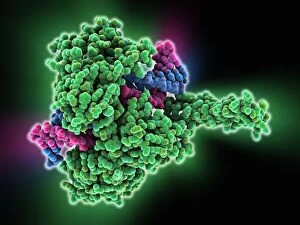
Type I topoisomerase bound to DNA. Molecular model showing a type I topoisomerase molecule (green) bound to a strand of DNA (deoxyribonucleic acid, pink and blue)

Background imageProteins Collection: Bombyx mori densovirus 1 capsid

Bombyx mori densovirus 1 capsid
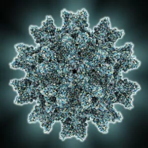
Bombyx mori densovirus 1 (BmDNV-1), molecular model. This virus infects crustaceans and insects, especially the silkworm (Bombyx mori)

Background imageProteins Collection: Methyladenine glycosylase bound to DNA

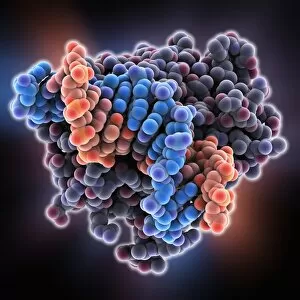
Methyladenine glycosylase bound to DNA. Computer model showing a molecule of human DNA-3-methyladenine glycosylase (purple) in complex with DNA (deoxyribonucleic acid, blue and orange)

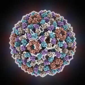
Background imageProteins Collection: Hepatitis B virus capsid, molcular model

Hepatitis B virus capsid, molcular model
Hepatitis B virus capsid, molecular model. This virus, transmitted through infected bodily fluids or blood, causes the disease hepatitis B in humans, leading to acute liver inflammation

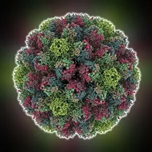
Background imageProteins Collection: Simian rotavirus capsid, molecular model

Simian rotavirus capsid, molecular model. This virus is named for its ability to infect the higher primates (simians). Rotaviruses, transmitted by faecal-oral contact

Background imageProteins Collection: HIV gp41 glycoprotein C014 / 0866

HIV gp41 glycoprotein C014 / 0866
HIV gp41 glycoprotein. Model showing the molecular structure of the gp41 protein from the HIV (human immunodeficiency virus) glycoprotein envelope

Background imageProteins Collection: Poliovirus type 3 capsid, molecular model

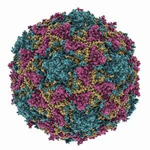
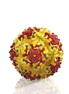
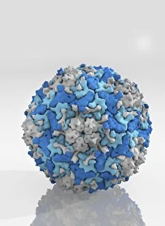
Poliovirus type 3 capsid, molecular model. This enterovirus causes poliomyelitis (polio) in humans, which affects the nervous system, sometimes leading to paralysis

Background imageProteins Collection: Transcription factor complexed with DNA C014 / 0869

Transcription factor complexed with DNA C014 / 0869
Transcription factor complexed with DNA. Computer model showing sterol regulatory element binding transcription factor 1 (SREBF1, horizontal) bound to a section of DNA (deoxyribonucleic acid)

Background imageProteins Collection: Zinc finger bound to DNA C014 / 0864

Zinc finger bound to DNA C014 / 0864
Zinc finger bound to DNA. Molecular model showing a zinc finger molecule bound to a strand of DNA (deoxyribonucleic acid)

Background imageProteins Collection: Beta-amyloid peptide molecule, artwork C014 / 2657

Beta-amyloid peptide molecule, artwork C014 / 2657
Molecular ribbon representation of the beta-amyloid peptide. The amyloid fibrillar form is the primary component of amyloid plaques found in the brains of Alzheimers disease patients

Background imageProteins Collection: Epstein-Barr virus protein bound to DNA C014 / 0875

Epstein-Barr virus protein bound to DNA C014 / 0875
Epstein-Barr virus protein bound to DNA. Computer model showing a molecule of Epstein-Barr nuclear antigen 1 (EBNA1) bound to a strand of DNA (deoxyribonucleic acid)

Background imageProteins Collection: HIV gp41 glycoprotein

HIV gp41 glycoprotein. Model showing the molecular structure of the gp41 protein from the HIV (human immunodeficiency virus) glycoprotein envelope

Background imageProteins Collection: ATP synthase molecule

ATP synthase molecule. Molecular model showing the structure of ATP synthase (ATPase) subunit C. ATPase is an important enzyme that provides energy for cells through the synthesis of adenosine

Background imageProteins Collection: Transcription factor complexed with DNA C014 / 0870

Transcription factor complexed with DNA C014 / 0870
Transcription factor complexed with DNA. Computer model showing a max protein (green) bound to a strand of DNA (deoxyribonucleic acid, pink)

Background imageProteins Collection: Nucleosome core particle bound to DNA C014 / 0872

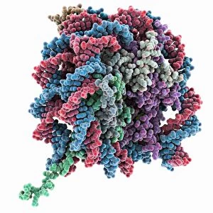
Nucleosome core particle bound to DNA C014 / 0872
Nucleosome core particle bound to DNA. Molecular model showing a nucleosome core particle (green and purple) bound to a strand of DNA (deoxyribonucleic acid, blue and red)

Background imageProteins Collection: Chaperonin protein complex C014 / 0873

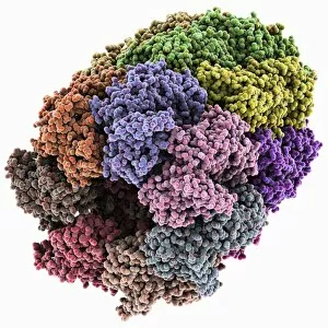
Chaperonin protein complex C014 / 0873
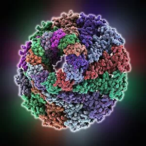
Chaperonin protein complex. Molecular model showing the structure of a GroEL/GroES/(ADP)7 chaperonin complex. Chaperonins are proteins that provide favourable conditions for the correct folding of

Background imageProteins Collection: Chaperonin protein complex

Chaperonin protein complex. Molecular model showing the structure of a GroEL/GroES/(ADP)7 chaperonin complex. Chaperonins are proteins that provide favourable conditions for the correct folding of

Background imageProteins Collection: Infectious bursal disease virus capsid

Infectious bursal disease virus capsid, molecular model. This avian virus infects the bursa of Fabricius (specialised bird immune organ) in young chickens, and can cause high mortality rates

Background imageProteins Collection: Animal cell organelles, artwork

Animal cell organelles, artwork
Animal cell organelles. Artwork showing the organelles in a eukaryotic cell. This is an animal cell. Structures include the nucleus (centre) which has a membrane with nuclear pores (purple)

Background imageProteins Collection: Bacteriorhodopsin protein

Bacteriorhodopsin protein. Molecular model showing the structure of bacteriorhodopsin (bR), a protein found in primitive micro-organisms known as Archaea. This protein acts as a proton pump

Background imageProteins Collection: Bacteriophage RNA, molecular model

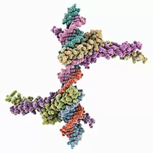
Bacteriophage RNA, molecular model
Bacteriophage RNA. Molecular model showing the structure of a loop of the genetic material RNA (ribonucleic acid) from a bacteriophage. Bacteriophages are viruses that infect bacteria

Background imageProteins Collection: Enterovirus particle C014 / 4900

Enterovirus particle C014 / 4900
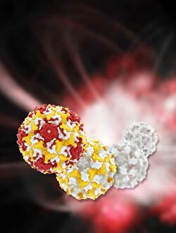
Enterovirus particle. Computer artwork of an enterovirus particle (virion), showing the structure of the capsid (outer shell)

Background imageProteins Collection: Bacteriophage connector protein

Bacteriophage connector protein. Molecular model showing the structure of a bacteriophage head-tail connector protein. Bacteriophages are viruses that infect bacteria

Background imageProteins Collection: Enterovirus capsid proteins structure C014 / 4897

Enterovirus capsid proteins structure C014 / 4897
Enterovirus capsid proteins structure. Computer artwork showing how the four component proteins (VP1 to VP4) of an enterovirus particle (virion) interlock to form the capsid (outer shell)

Background imageProteins Collection: Enterovirus capsid proteins structure C014 / 4896

Enterovirus capsid proteins structure C014 / 4896
Enterovirus capsid proteins structure. Computer artwork showing how the four component proteins (VP1 to VP4) of an enterovirus particle (virion) interlock to form the capsid (outer shell)

Background imageProteins Collection: MscS ion channel protein structure

MscS ion channel protein structure. Molecular model showing the protein structure of a Mechanosensitive Channel of Small Conductance (MscS) from an Escherichia coli bacterium

Background imageProteins Collection: Bacteriophage capsid protein shell

Bacteriophage capsid protein shell. Molecular model showing the partial shell structure of a bacteriophage capsid based on one of its coat proteins

Background imageProteins Collection: Alemtuzumab antibody molecule

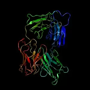
Alemtuzumab antibody molecule. Computer model showing the molecular structure of the monoclonal antibody and cancer drug alemtuzumab

Background imageProteins Collection: Enterovirus particle C014 / 4898

Enterovirus particle C014 / 4898
Enterovirus particle. Computer artwork of an enterovirus particle (virion), showing the structure of the capsid (outer shell)

Background imageProteins Collection: Human poliovirus, molecular model

Human poliovirus, molecular model
Human poliovirus particle. Computer model of the capsid of the human poliovirus. The capsid is a protein coat that encloses the viruss genetic information (genome), stored as RNA (ribonucleic acid)


All Professionally Made to Order for Quick Shipping

Proteins: The Building Blocks of Life From the intricate network of nerve and glial cells to the mesmerizing patterns seen under a light micrograph, proteins play an essential role in every aspect of our existence, and are like the conductors of our body's symphony, orchestrating vital processes that keep us alive and functioning. Take, for example, an anaesthetic inhibiting an ion channel C015 / 6718. Proteins act as gatekeepers, controlling what enters or exits our cells. In this case, they regulate the flow of ions necessary for transmitting nerve signals and maintaining proper cell function. But proteins don't just govern our internal workings; they also interact with external threats such as the avian flu virus. These microscopic invaders hijack host cells using their own protein machinery to replicate themselves. Understanding these interactions is crucial in developing effective treatments against viral infections. While some proteins protect us from harm, others contribute to overall well-being through a balanced diet. Our bodies require various types found in different foods to ensure optimal health and nutrition. The secondary structure is truly a work of art—a complex folding pattern that determines their shape and function. Artists have captured this beauty through stunning artwork showcasing these intricate molecular structures. One such structure is the nucleosome molecule—an elegant arrangement where DNA wraps around protein spools called histones—forming compact units within chromosomes. This organization allows efficient storage and retrieval of genetic information during cell division or gene expression. Antibodies are another remarkable class depicted in captivating artwork. These specialized molecules recognize foreign substances like bacteria or viruses and neutralize them by binding tightly to specific targets on their surface—an extraordinary defense mechanism employed by our immune system. Speaking of bacteria, their ribosomes serve as factories producing new proteins based on instructions encoded in DNA—the blueprint for life itself. Understanding bacterial ribosomes has led to groundbreaking discoveries in antibiotic development, combating infectious diseases that threaten human health.