
Proteomics Collection
Proteomics, the study of proteins and their functions within an organism, is a fascinating field that unravels the intricate workings of life


All Professionally Made to Order for Quick Shipping
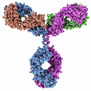
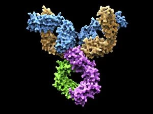
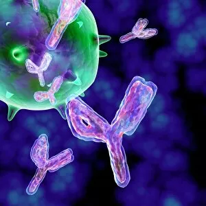
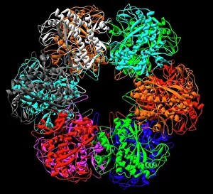
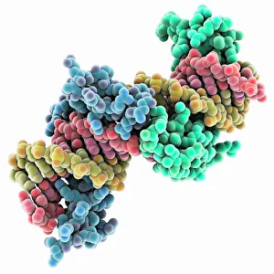
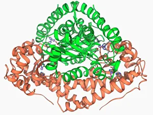
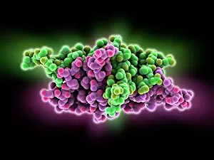
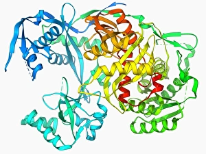
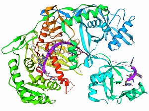
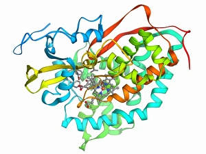
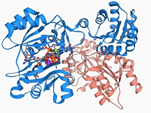
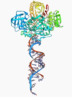
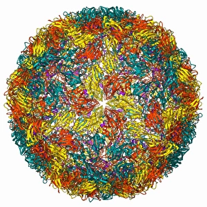
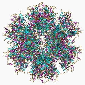
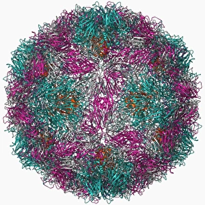
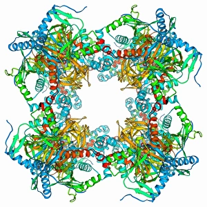
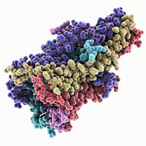
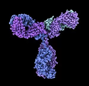
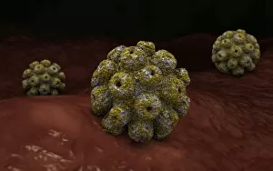
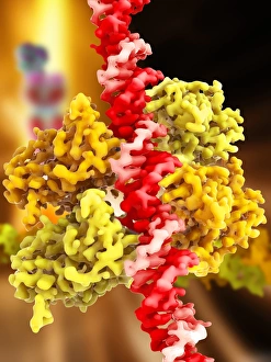
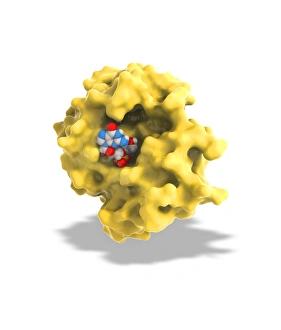
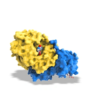
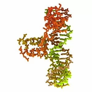
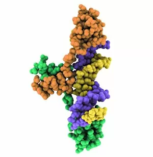
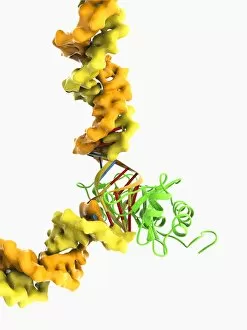
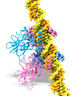
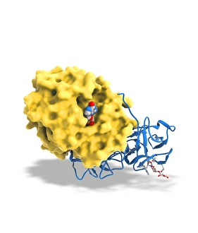
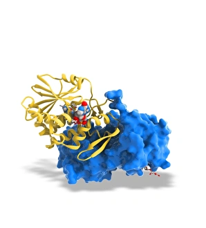
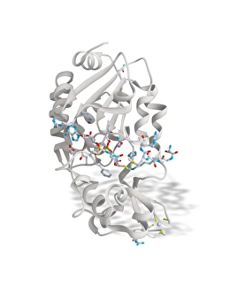
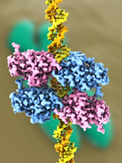
Proteomics, the study of proteins and their functions within an organism, is a fascinating field that unravels the intricate workings of life. From anaesthetics inhibiting ion channels to immunoglobulin G antibody molecules, proteomics delves into the molecular mechanisms that shape our existence. In the realm of brain research, scientists explore how proteins influence cognition and behavior. They investigate DNA nucleosomes' structure and function, unraveling their role in gene regulation. Antibodies take center stage as artwork showcases their diverse forms and crucial role in immune defense. Zinc fingers bound to a DNA strand highlight protein-DNA interactions critical for genetic processes. Meanwhile, manganese superoxide dismutase enzyme aids in protecting cells from oxidative stress. The SARS coronavirus protein becomes a subject of intense scrutiny as researchers strive to understand its pathogenicity. Cytochrome b5 molecule reveals insights into electron transfer reactions within cells while glutamine synthetase enzyme plays a vital role in nitrogen metabolism. Lastly, RNA-editing enzymes offer potential therapeutic targets for various diseases with their ability to modify genetic information at the RNA level. Through proteomics, we unlock nature's secrets one protein at a time - deciphering their structures, unraveling their functions, and ultimately enhancing our understanding of life itself.