
Pulmonary Collection
"Pulmonary: Unveiling the Intricacies of our Vital Breath" Discover the wonders of the pulmonary system through a captivating journey


All Professionally Made to Order for Quick Shipping
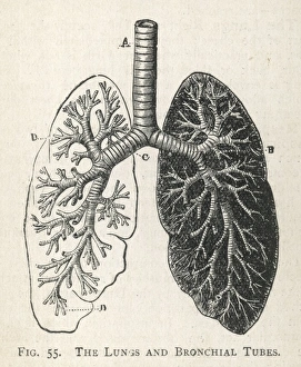
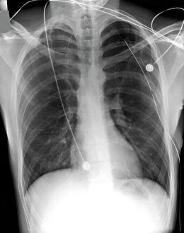
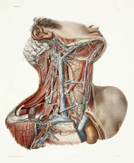
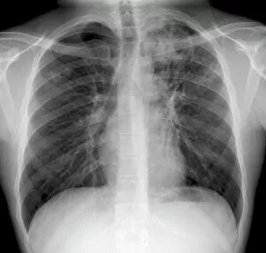
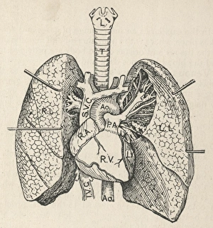
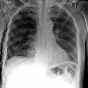
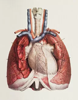
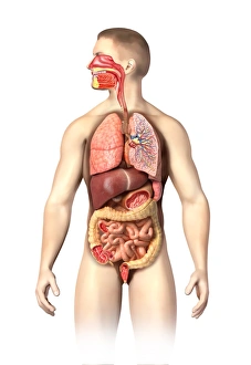
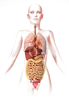
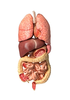
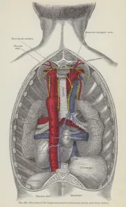
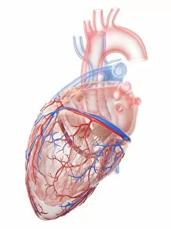
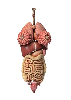
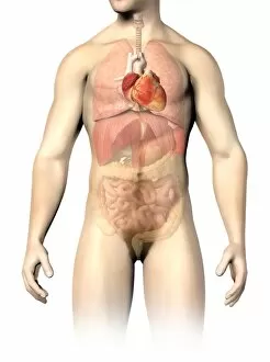
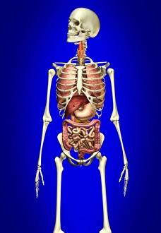
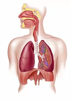
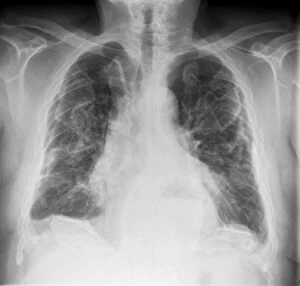
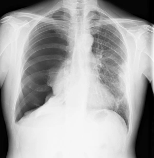
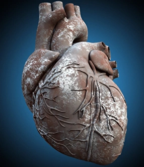
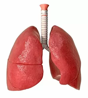
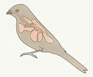
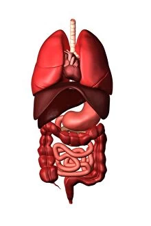
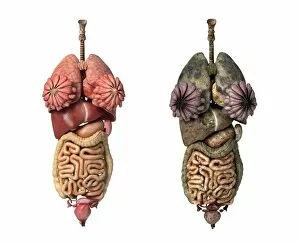
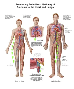
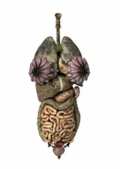
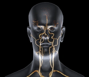
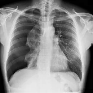
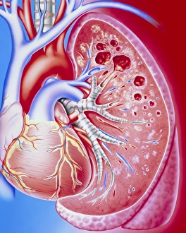
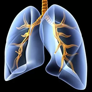
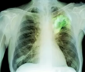
"Pulmonary: Unveiling the Intricacies of our Vital Breath" Discover the wonders of the pulmonary system through a captivating journey. Dive into a world where life-sustaining breaths take center stage, as we explore various aspects that define our respiratory health. Embark on this educational voyage with a detailed diagram showcasing the intricate structure of lungs and bronchial tubes. Marvel at their complexity, understanding how they facilitate oxygen exchange within our bodies. Witness medical advancements in pneumothorax treatment through X-ray imagery, offering hope to those affected by this condition. Delve into neck anatomy studies, unraveling the secrets hidden beneath our skin while appreciating 19th Century artwork depicting these discoveries. Uncover the devastating impact of tuberculosis through haunting X-ray images that reveal its presence within afflicted lungs. Gain insight into how interconnected systems work together harmoniously with an enlightening diagram illustrating the heart, lungs, and windpipe. Experience tension pneumothorax's urgency via gripping X-rays that depict its severity and prompt intervention requirements. Grasp the symbiotic relationship between heart and lungs; two vital organs intricately linked for optimal functioning. Explore cystic fibrosis' challenges faced by patients daily; understand their resilience amidst adversity. Learn about Konstantin Buteyko's groundbreaking contributions as a Soviet doctor revolutionizing respiratory therapy techniques. Marvel at abdominal arteries' inner workings revealed in an illuminating X-ray image (P206 / 0309), highlighting their significance in maintaining overall health. Discover Hantavirus' impact on pulmonary health – raising awareness about potential risks lurking in nature's realm. Reflect upon "HOCHE DIES AT WETZLAR, " reminding us of historical milestones shaping modern medicine's understanding diseases throughout time. Join us on this extraordinary expedition exploring every facet of "pulmonary. " Let curiosity guide you as we unravel mysteries surrounding respiration - an awe-inspiring process essential to our very existence.