
Radiography Collection
Radiography, the art of capturing hidden wonders through the lens of X-rays, unveils a captivating world beyond our naked eyes


All Professionally Made to Order for Quick Shipping
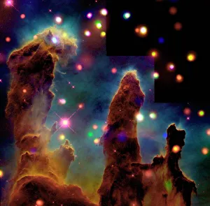
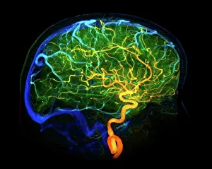
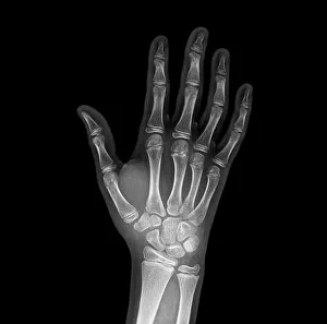
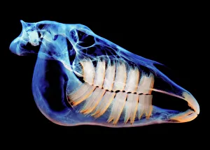
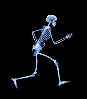
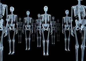
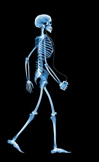
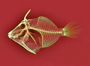
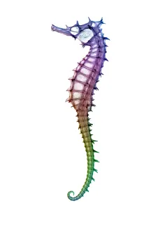
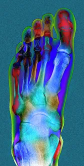
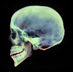
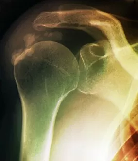
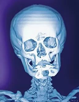
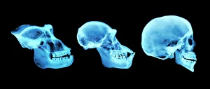
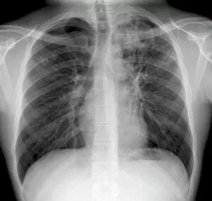
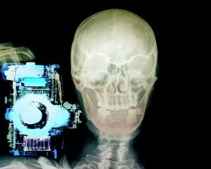
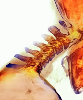
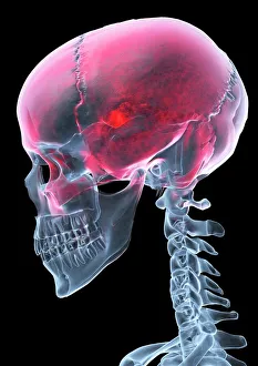
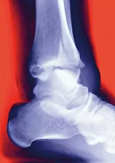
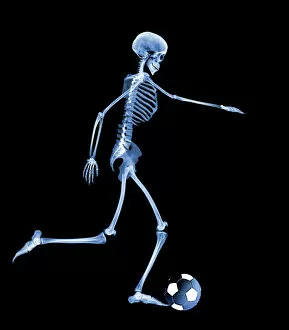
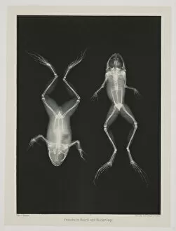
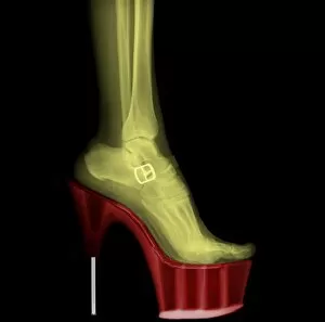
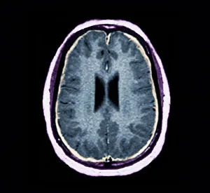
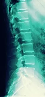
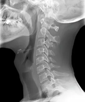
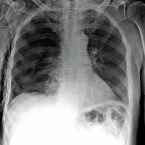
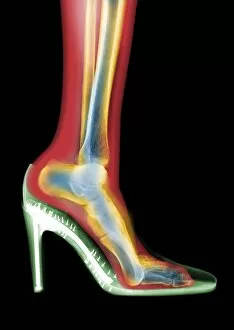
Radiography, the art of capturing hidden wonders through the lens of X-rays, unveils a captivating world beyond our naked eyes. Like the awe-inspiring "Pillars of Creation" in space, radiography allows us to explore intricate details within our own bodies and beyond. Delving into the depths of human anatomy, it reveals the delicate network of brain blood vessels with a mesmerizing 3D angiogram from 1981. A broken wrist bone is laid bare by an X-ray, showcasing both fragility and resilience in one image. Beyond humans, radiography takes us on a journey through nature's marvels. The majestic skull of a horse reminds us of their grace and strength. Meanwhile, even skeletons find joy as we witness one playing rugby - an unexpected twist that brings humor to this scientific art form. But radiography doesn't stop at living beings; it delves into dental health too. A panoramic dental X-ray captures every tooth's story while revealing potential issues lurking beneath the surface. Even marine life isn't spared from its gaze: an ethereal lobster appears ghostly yet beautiful when seen through an X-ray lens. The fusion between technology and humanity becomes evident as we encounter a person holding a camera captured in an X-ray frame – reminding us that even those behind the lens are not exempt from exploration. Intriguingly surreal images emerge as skeletons engage in activities typically associated with life: drinking or engaging in sports like rugby. These playful depictions remind us that there is always more than meets the eye when it comes to our inner structures. Radiography transcends earthly boundaries too; it ventures into outer space where composite images reveal spiral galaxy M81's cosmic dance among stars – merging science and art seamlessly together. From triggerfish to seahorses, radiography exposes intricate skeletal structures unseen under normal circumstances—reminding us that beauty exists even within what may seem ordinary or overlooked.