Fraud Blocker Skip to main content

Re Production Collection (page 9)

Exploring the wonders of reproduction through time and nature

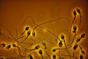
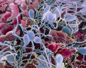
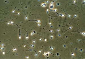
Background imageRe Production Collection: LM of normal human sperm

LM of normal human sperm

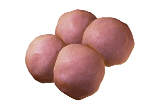
Background imageRe Production Collection: 4-cell embryo embryo, artwork

4-cell embryo embryo, artwork
4-cell embryo. Image 3 of 4. Computer model representing 4 daughter cells. The development of an embryo is called embryogenesis

Background imageRe Production Collection: LM of human sperm fertilising an egg

LM of human sperm fertilising an egg
Sperm fertilising egg. Light micrograph of a human egg with sperm at its surface attempting to penetrate and fertilise it

Background imageRe Production Collection: Egg cell, SEM

Egg cell, SEM
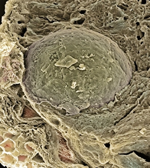
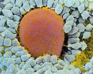
Egg cell. Coloured scanning electron micrograph (SEM) of a freeze-fracture of a developing egg cell in a secondary follicle of the ovary. At centre is the rounded egg cell or secondary oocyte (red)

Background imageRe Production Collection: Coloured SEM of egg cell in secondary follicle

Coloured SEM of egg cell in secondary follicle

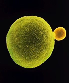
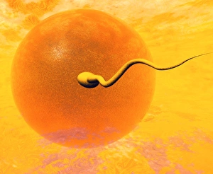
Background imageRe Production Collection: Sperm and egg

Sperm and egg
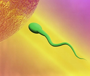
Fertilisation. Computer artwork of a human sperm approaching an egg (ovum, top left) just before the process of fertilisation

Background imageRe Production Collection: Blood vessels in a frog ovary, SEM

Blood vessels in a frog ovary, SEM
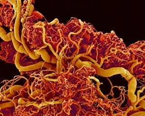
Blood vessels in the ovary of a female frog, coloured scanning electron micrograph (SEM). Frogs produce hundreds of eggs, which are expelled and fertilised in the water

Background imageRe Production Collection: Human embryo for sale

Human embryo for sale, conceptual computer artwork. Human embryos are fertilised eggs in their earliest stage of development; less than 8 weeks old

Background imageRe Production Collection: False-colour SEM of sperm in the uterine cavity

False-colour SEM of sperm in the uterine cavity

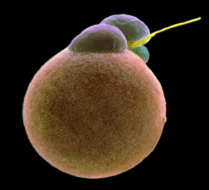
Background imageRe Production Collection: Coloured SEM of sperm fertilising an egg

Coloured SEM of sperm fertilising an egg
Coloured scanning electron micrograph of sperm (yellow) fertilising an egg cell (round and pink). At top centre three polar bodies (purple and blue) are seen

Background imageRe Production Collection: Frog embryo, light micrograph

Frog embryo, light micrograph
Frog embryo. Light micrograph of a frog embryo at the blastula stage. This stage in the embryos development is produced by the cleavage (cell division) of a fertilised ovum (egg cell)

Background imageRe Production Collection: Gene transfer in fertilisation

Gene transfer in fertilisation

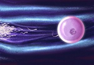
Background imageRe Production Collection: Computer artwork of human sperm fertilising an egg

Computer artwork of human sperm fertilising an egg
Sperm fertilising egg. Computer illustration of sperm cells (white) approaching a human egg (pink), before one of the sperm fertilises it

Background imageRe Production Collection: Coloured SEM of sperm on egg during fertilisation

Coloured SEM of sperm on egg during fertilisation
Fertilisation. Coloured scanning electron micro- graph (SEM) of sperm on the surface of a human egg (ovum) during fertilisation. The egg has a spongy protective surface

Background imageRe Production Collection: Ovulation, artwork

Ovulation, artwork
Ovulation. Computer artwork of an egg (yellow) being released from an ovary (bottom), a process known as ovulation. The egg is being released into a fallopian tube (top)

Background imageRe Production Collection: False-colour SEM of the ovary surface at ovulation

False-colour SEM of the ovary surface at ovulation

Background imageRe Production Collection: Coloured LM of a fertilised human egg 24 hours old

Coloured LM of a fertilised human egg 24 hours old

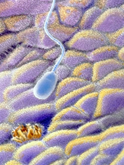
Background imageRe Production Collection: False-colour SEM of a spermatozoon on uterus wall

False-colour SEM of a spermatozoon on uterus wall
A spermatozoon on the uterine mucosa. False-colour scanning electron micrograph of a spermatozoon floating over the endometrium, the internal wall of the uterus

Background imageRe Production Collection: False-colour SEM of secondary oocyte & polar body

False-colour SEM of secondary oocyte & polar body
Human egg. False-colour scanning electron micrograph of a human egg (primary oocyte) at the end of the first meiotic division, occurring just before ovulation

Background imageRe Production Collection: SEM of ovulation; release of egg

SEM of ovulation; release of egg

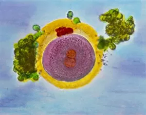
Background imageRe Production Collection: Egg fertilisation

Egg fertilisation
False-colour transmission electron micrograph (TEM) of a single human ovum (egg) surrounded by sperm. The egg has been coloured orange, and the numerous sperm appear rougly spherical in this section

Background imageRe Production Collection: Sperm fertilising egg

Sperm fertilising egg

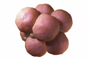
Background imageRe Production Collection: Multi-celled embryo, artwork

Multi-celled embryo, artwork
Multi-celled embryo. Image 4 of 4. Computer model representing a cluster of daughter cells. The development of an embryo is called embryogenesis

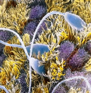
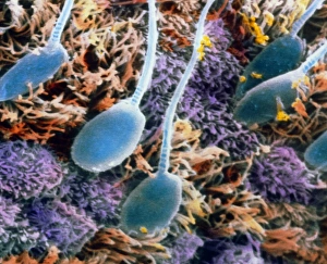
Background imageRe Production Collection: False-colour SEM of sperm on uterine surface

False-colour SEM of sperm on uterine surface
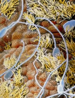
Capacitation of spermatozoa. False-colour scanning electron micrograph of a group of spermatozoa (turquoise) floating over the endometrium, the internal wall of the uterus

Background imageRe Production Collection: Artwork of sperm and egg just before fertilisation

Artwork of sperm and egg just before fertilisation

Background imageRe Production Collection: False-colour SEM of sperm on a human egg

False-colour SEM of sperm on a human egg
Fertilisation. False-colour scanning electron micrograph of sperm (blue) on a human egg. In order to fertilise the egg a spermatozoon must penetrate the thick layer of follicular cells (pink)

Background imageRe Production Collection: Single-celled embryo, artwork

Single-celled embryo, artwork
Single-celled embryo. Image 1 of 4. Computer model representing a single cell. The development of an embryo is called embryogenesis

Background imageRe Production Collection: Human fertilisation

Human fertilisation
Sperm fertilising egg. Computer illustration of sperm cells (at right) approaching a human egg (at left). One of the sperm is fertilising it

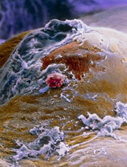
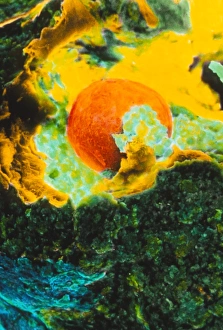
Background imageRe Production Collection: Coloured SEM showing the ovulation process

Coloured SEM showing the ovulation process
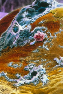
Ovulation. Coloured scanning electron micrograph of the ovulation process. The egg (red at centre) has ruptured the external surface of the ovary (dark green), known as the germinal epithelium

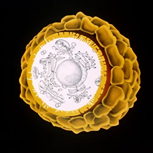
Background imageRe Production Collection: Artwork of a human egg cell

Artwork of a human egg cell in the primordial follicle phase which occurs after seven months of fetal development. The egg cell is surrounded by a thick layer of follicular cells known as zona

Background imageRe Production Collection: Light micrograph of human sperm cells

Light micrograph of human sperm cells

Background imageRe Production Collection: 2-cell embryo embryo, artwork

2-cell embryo embryo, artwork
2-cell embryo. Image 2 of 4. Computer model representing 2 daughter cells. The development of an embryo is called embryogenesis

Background imageRe Production Collection: False-colour SEM of human egg after ovulation

False-colour SEM of human egg after ovulation
Ovarys surface at ovulation. False-colour scanning electron micrograph of the surface of the ovary about one hour after ovulation

Background imageRe Production Collection: Sperm in the testis, TEM

Sperm in the testis, TEM
Sperm cells. Coloured transmission electron micrograph (TEM) of a section through the testis, showing sperm cells. The heads of the sperm, which contain the genetic material, are red

Background imageRe Production Collection: False-colour SEM of human sperm in the uterus

False-colour SEM of human sperm in the uterus

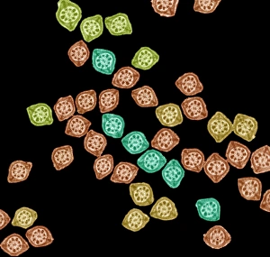
Background imageRe Production Collection: Sperm tails, TEM

Sperm tails, TEM
Sperm tails. Coloured transmission electron micrograph (TEM) of cross sections through sperm tails. A sperms tails ability to move is partly due to the arrangement of microtubules

Background imageRe Production Collection: Human sperm

Human sperm

Background imageRe Production Collection: Sperm cell, computer artwork

Sperm cell, computer artwork
Sperm cell. Computer artwork of a human sperm cell, showing its inner structure. The sperm head (orange) carries genetic material

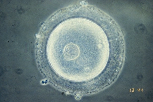
Background imageRe Production Collection: LM of immature human oocyte with germinal vesicle

LM of immature human oocyte with germinal vesicle
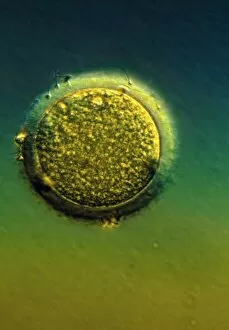
Light micrograph of an immature human oocyte (the female egg cell) containing a germinal body, the large cell nucleus. Oocytes are formed in the ovaries

Background imageRe Production Collection: Dividing cell

Dividing cell. Differential interference contrast (DIC) light micrograph of a cell (lower left) in the metaphase stage of mitosis (cell division). The cells nuclei are stained with fluorescent dye

Background imageRe Production Collection: Gender determination

Gender determination, computer artwork. Gender is determined by the sex chromosomes, X and Y. Females have 2 X chromosomes and men an X and a Y chromosome

Background imageRe Production Collection: False-colour SEM of sperm in uterus

False-colour SEM of sperm in uterus
Sperm in uterus. False-colour scanning electron micrograph of a human sperm on the inner surface of the uterus, the endometrium

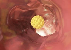
Background imageRe Production Collection: Embryo in a fallopian tube, artwork

Embryo in a fallopian tube, artwork
Embryo inside a fallopian tube. Computer artwork representing an embryo (yellow) in the early stages of development inside a fallopian tube. Fallopian tubes lead from the ovaries to the uterus

Background imageRe Production Collection: False-colour SEM of human egg surrounded by sperm

False-colour SEM of human egg surrounded by sperm

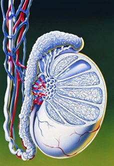
Background imageRe Production Collection: Artwork of section through human testis

Artwork of section through human testis
Illustration of a section through a single human testis, one of a pair of male sex organs that are the site of production of spermatozoa (spermatogenesis) and androgen, the male sex hormone

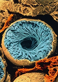
Background imageRe Production Collection: Colour SEM of seminiferous tubule of the testis

Colour SEM of seminiferous tubule of the testis
Seminiferous tubule. Coloured Scanning Electron Micrograph (SEM) of a sectioned seminiferous tubule, the site of sperm production in the human testis

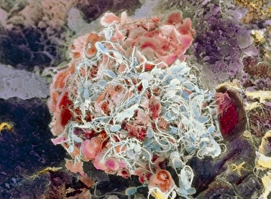
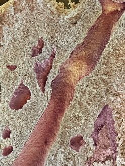
Background imageRe Production Collection: Ovarian tissue, SEM

Ovarian tissue, SEM
Ovarian tissue. Coloured scanning electron micrograph (SEM) of a section through the medulla of an ovary showing blood vessels (pink)

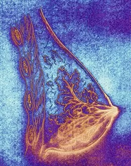
Background imageRe Production Collection: Artwork of a healthy breast

Artwork of a healthy breast
Healthy breast. Artwork of a normal female breast, seen in side view. The nipple is seen at lower right, with the glandular lobules which secrete milk seen as leaf-like structures within the breast


All Professionally Made to Order for Quick Shipping

Exploring the wonders of reproduction through time and nature. 🌍🔬 From the intricate Catalan Atlas of the 14th century to Da Vinci's innovative crossbow, humans have always sought ways to understand and enhance reproduction. Witness the ancient mating rituals of Tyrannosaurus rex dinosaurs captured in fossils, or marvel at the microscopic beauty of a uterus lining during menstruation under an SEM microscope. Delve into the miracle of life with illustrations showcasing the human placenta or observe English oak acorns as they prepare for their own reproductive journey. Discover nature's secrets with stunning images of geranium anthers and dahlia flower pollen magnified by SEM technology. Follow maple seed flight paths as they embark on their quest for new beginnings, just like Robert Louis Stevenson once wrote about in his captivating works on reproduction. Dive deep into marine life with sea cucumbers, fascinating creatures that possess unique methods of reproduction. And finally, explore the intricate world of plant fertility with mesmerizing SEM images capturing pollen grains in all their glory. Join us on this incredible journey through time and nature as we unravel the mysteries behind 'reproduction'.