
Receptor Collection
"Exploring the Intricate World of Receptors: From Anaesthetic Inhibition to Brain Protein Research" Receptors play a vital role in various aspects of our lives


All Professionally Made to Order for Quick Shipping
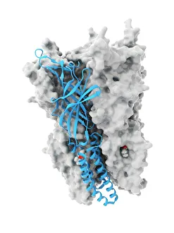
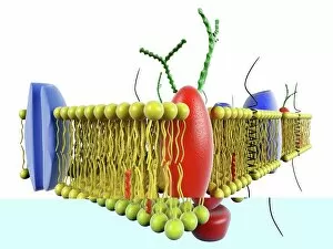
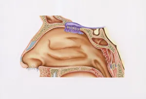
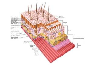
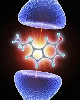
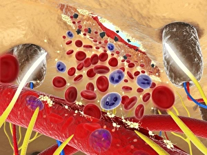
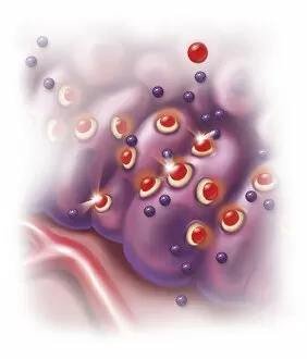
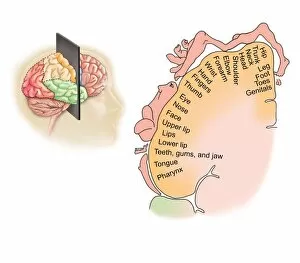
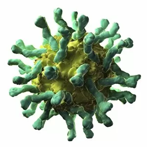
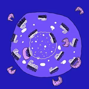
"Exploring the Intricate World of Receptors: From Anaesthetic Inhibition to Brain Protein Research" Receptors play a vital role in various aspects of our lives, from the intricate workings of our brain to the sensory experiences that shape our perception. One fascinating example is how anaesthetics inhibit ion channels (C015 / 6718), providing relief during medical procedures. In the realm of brain protein research, receptors take center stage as scientists delve into understanding their complex interactions and functions. These proteins act as gatekeepers, regulating signals within neural networks and shaping our cognitive abilities. Art meets science when we examine receptor activity at the cell membrane (artwork C013 / 7467). This captivating image showcases the delicate dance between molecules that occurs on this crucial boundary, influencing cellular communication and signaling pathways. Our eyes are also home to remarkable receptors known as rod and cone cells (SEM C014 / 4866). These microscopic structures enable us to perceive light and colors, painting vivid pictures of the world around us. Delving into history, even amidst tumultuous times like the Spanish Civil War (1936-1939), "El receptor de" becomes an emblematic phrase symbolizing resilience and hope for those longing for connection amid chaos. Moving beyond sight, olfaction takes center stage with smell receptors found in nasal epithelium (cross-section illustration). These specialized receptors allow us to savor fragrances while unraveling memories associated with scents—a gateway to nostalgia itself. Stepping back in time technologically, we encounter a radio-receiver with a superheterodyne circuit featuring only four valves. This iconic device demonstrates how early engineers harnessed receptor principles to bring music and news into homes worldwide. Shifting gears towards anatomy, exploring human skin reveals its intricate layers through conceptual images or detailed diagrams. Understanding these layers helps comprehend how receptors embedded within transmit sensations such as touch or temperature—our body's first line of defense.