
Red Blood Cells Collection
"Exploring the Intricate World of Red Blood Cells: From Menstruation to Malaria" Uterus lining during menstruation


All Professionally Made to Order for Quick Shipping
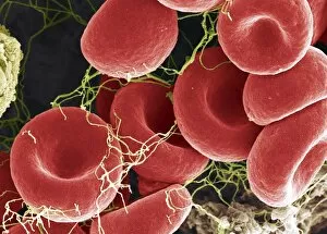
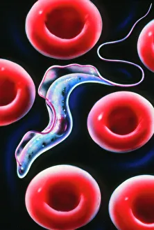
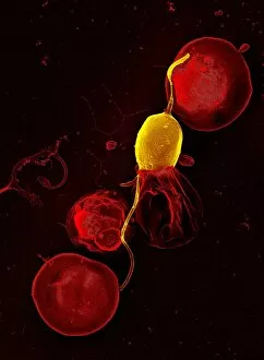
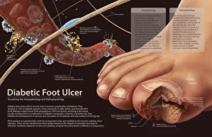
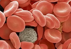
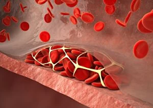
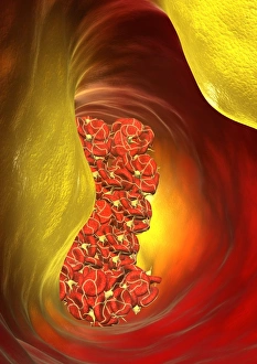
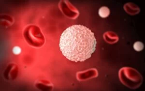
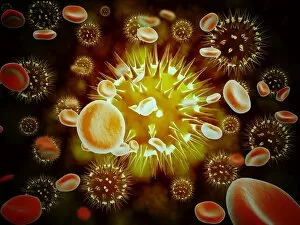
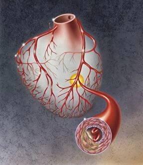
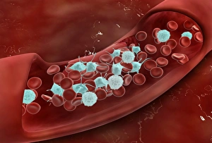
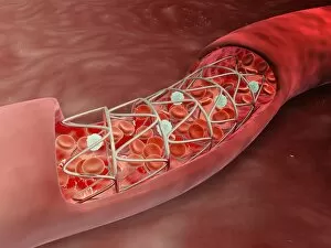
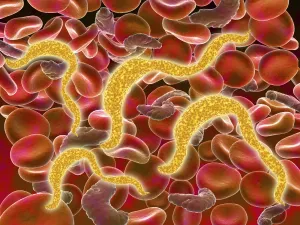
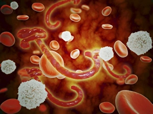
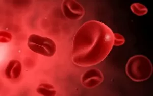
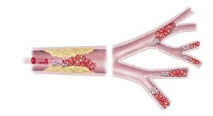
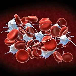
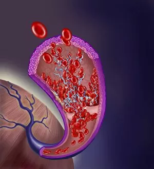
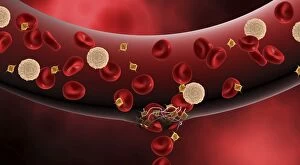
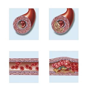
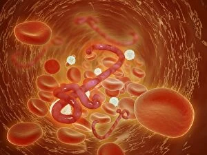
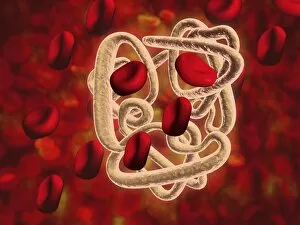
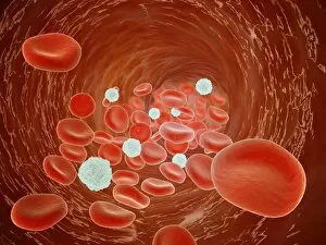
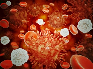
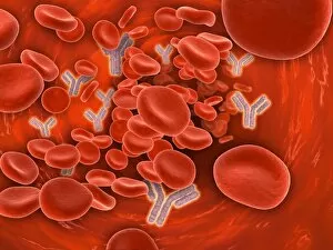
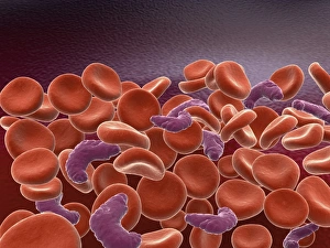
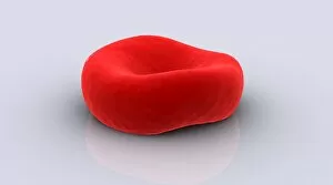
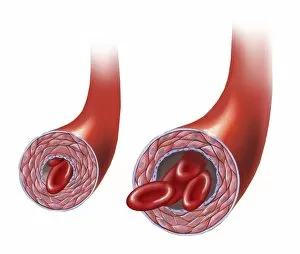
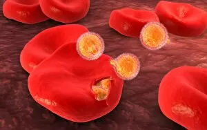
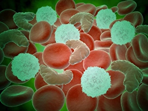
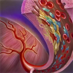
"Exploring the Intricate World of Red Blood Cells: From Menstruation to Malaria" Uterus lining during menstruation, SEM: Witness the fascinating transformation of the uterus lining during menstruation as red blood cells play a crucial role in this natural process. Red and white blood cells, SEM: Delve into the microscopic realm where red and white blood cells coexist, working together to maintain our body's health and vitality. Blood clot, SEM C016 / 9747: Uncover the intricate structure of a blood clot under scanning electron microscopy, highlighting how red blood cells contribute to its formation. Red blood cells, SEM: Marvel at the mesmerizing beauty of individual red blood cells captured through scanning electron microscopy, showcasing their unique shape and function. Sleeping sickness parasite: Explore how red blood cells become unwitting hosts for parasites causing sleeping sickness, shedding light on this devastating disease's impact on human health. Mouse malaria parasite, SEM: Dive into the world of malaria as we examine mouse malaria parasites residing within red blood cells using scanning electron microscopy techniques. Malaria parasites, TEM: Peer deep inside infected red blood cells with transmission electron microscopy to unravel the complex life cycle and pathogenicity of malaria parasites. Artery SEM: Take a closer look at an artery's inner wall under scanning electron microscopy to understand how healthy red blood cell flow is vital for cardiovascular well-being. Deep vein thrombus: Discover how disrupted circulation can lead to dangerous deep vein thrombosis as clots form within veins due to abnormal interactions between platelets and red blood cells. Histopathology and pathophysiology of diabetic foot ulcers: Gain insights into diabetes-related complications by examining histopathological changes in foot ulcers caused by impaired microcirculation involving damaged capillaries and compromised oxygen supply from reduced numbers or functionality of red blood cells.