
Regulatory Collection
"Exploring the Intricacies of Regulatory Systems: From Pancreas Anatomy to Artwork" Delving into the intricate world systems


All Professionally Made to Order for Quick Shipping
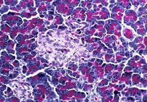
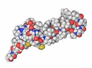
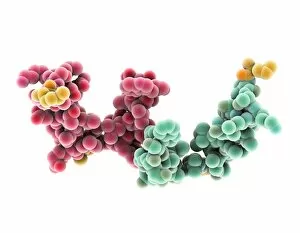
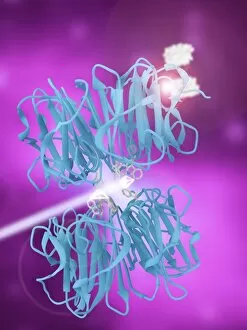
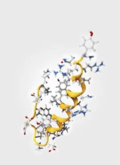
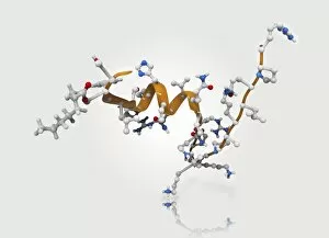
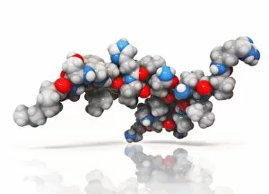
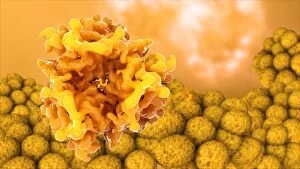
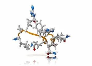
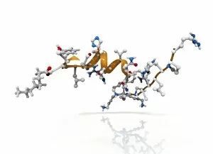
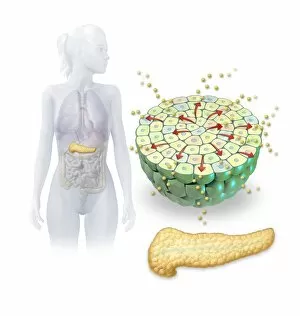
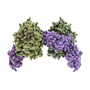
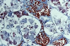
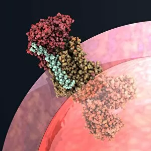
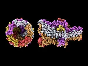
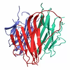
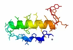
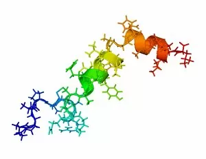
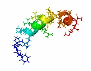
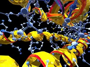
"Exploring the Intricacies of Regulatory Systems: From Pancreas Anatomy to Artwork" Delving into the intricate world systems, we uncover the fascinating anatomy of the pancreas and its role in maintaining homeostasis. Through a scanning electron microscope (SEM), English oak leaf pores reveal their hidden secrets, shedding light on how regulation occurs at a microscopic level. The pancreatic islet of Langerhans takes center stage as we explore its vital function in regulating blood sugar levels and insulin production. French lavender leaf pores captured under SEM unveil nature's own regulatory mechanisms, showcasing the beauty within even the tiniest details. Artistic renditions bring Islets of Langerhans cells to life, highlighting their crucial role in coordinating hormonal responses for optimal bodily regulation. "Taking the Census, 1854" by an unknown artist reminds us that data collection has long been essential for understanding populations and implementing effective regulations. Zooming into molecular scale, we examine the parathyroid hormone molecule and its pivotal role in calcium regulation within our bodies' complex systems. The Council of Europe building stands tall amidst flags representing member nations - a symbol of international cooperation towards harmonizing regulatory frameworks across borders. Unveiling another molecular player, cAMP-dependent protein kinase molecule F006/9728 emerges as a key regulator involved in numerous cellular processes throughout our body. Sex hormone-binding globulin molecule F006/9281 comes into focus as we unravel how it modulates sex hormones' availability and influences various physiological functions. A glimpse through transmission electron microscopy (TEM) reveals intricate structures within the anterior pituitary gland - an organ responsible for producing several important hormones involved in systemic regulation. Hepatocyte nuclear factor molecule C015/7699 takes center stage as we explore its critical involvement in liver development, metabolism, and regulatory processes.