
Replicating Collection
"Unveiling the Art of Replication: From Monks Copying Manuscripts to Budding HIV Particles" In the ancient halls of monasteries


All Professionally Made to Order for Quick Shipping
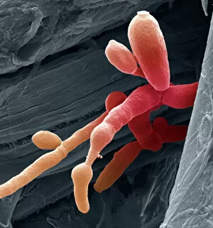
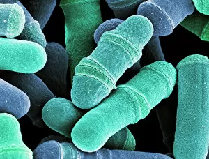
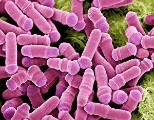
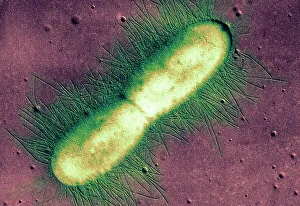
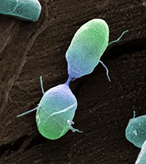
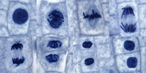
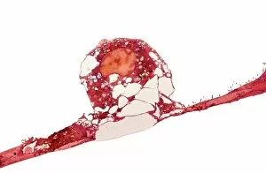
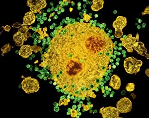
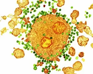
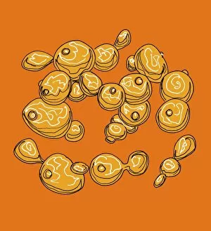
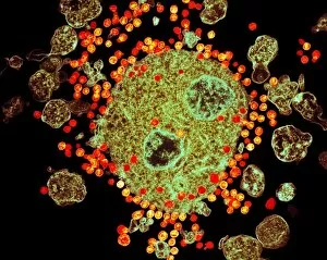
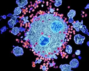
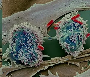
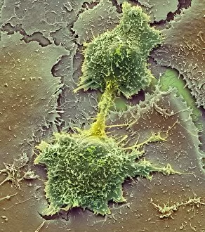
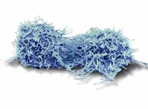
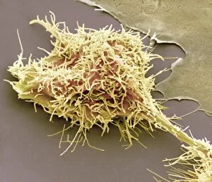
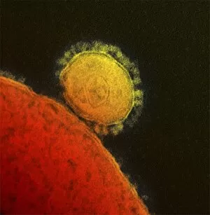
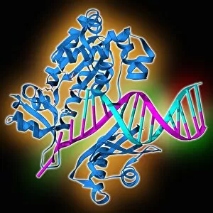
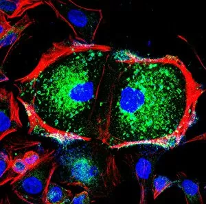
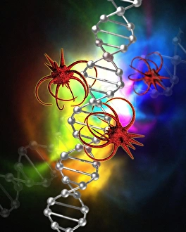
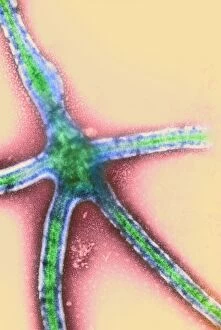
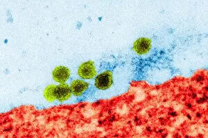
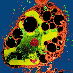
"Unveiling the Art of Replication: From Monks Copying Manuscripts to Budding HIV Particles" In the ancient halls of monasteries, dedicated monks meticulously replicate manuscripts, preserving knowledge for generations to come. (Monks Copying Manuscript) Underneath our very noses, a microscopic world thrives with replication as Candida fungus multiplies and spreads its presence. (Candida fungus, SEM) Witness the mesmerizing dance of yeast cells dividing, their intricate process captured in stunning detail under the scanning electron microscope. (Dividing yeast cells, SEM) The relentless nature of life continues as herpes virus replicates within host cells, perpetuating its existence and causing discomfort. (Herpes virus replicating) Delving deeper into unseen realms reveals intestinal protozoan parasites undergoing replication - an astonishing sight through the transmission electron microscope. (Intestinal protozoan parasites, TEM) Behold the striking image of Vesicular stomatitis virus particles multiplying relentlessly inside host cells - a testament to their survival instincts. (Vesicular stomatitis virus, TEM) Peering into the microcosm once again uncovers E. coli bacterium replicating with remarkable precision and efficiency - an awe-inspiring feat captured by the transmission electron microscope. (E. coli bacterium, TEM) Salmonella bacterium divides before our eyes in this captivating scanning electron microscopy image – showcasing how they propagate their lineage through division. (Salmonella bacterium dividing, SEM) Cell division unfolds like a delicate ballet on a grand stage; witness this breathtaking spectacle through high-resolution scanning electron microscopy imagery. (Cell division, SEM) Plant cell mitosis comes alive under light micrography's gentle touch – revealing nature's elegant way of creating new life from existing ones. (Plant cell mitosis, light micrograph) Budding HIV particles emerge from infected cells like tiny alien spacecrafts ready to infect anew - a hauntingly beautiful sight captured by scanning electron microscopy.