Fraud Blocker Skip to main content

Respiratory Collection

"The Marvels of the Respiratory System: Exploring the Gateway to Life" Diagram of the Lungs and Bronchial Tubes

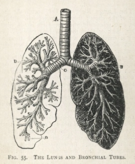
Background imageRespiratory Collection: Diagram of the lungs and bronchial tubes

Diagram of the lungs and bronchial tubes
A diagram of the lungs and bronchial tubes, with a section of the windpipe

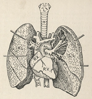
Background imageRespiratory Collection: Diagram of the heart, lungs and windpipe

Diagram of the heart, lungs and windpipe

Background imageRespiratory Collection: Deer anatomy, artwork

Deer anatomy, artwork
Deer anatomy. Computer artwork showing the internal anatomy of a deer (family Cervidae). Anatomical features shown here include the skeleton, lungs, intestines, bladder

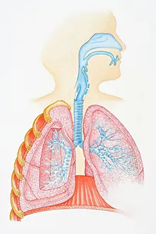
Background imageRespiratory Collection: Illustration of human respiratory system showing oral cavity, and nasal cavity, larynx, trachea

Illustration of human respiratory system showing oral cavity, and nasal cavity, larynx, trachea, bronchus, and lungs

Background imageRespiratory Collection: Konstantin Buteyko, Soviet doctor

Konstantin Buteyko, Soviet doctor
Konstantin Pavlovich Buteyko (1923-2003), Soviet medical doctor and researcher. In the 1950s, Buteyko, a Ukrainian, developed a theory on the effects of deep breathing (hyperventilation)

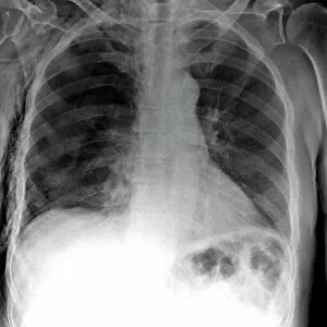
Background imageRespiratory Collection: Tension pneumothorax, X-ray

Tension pneumothorax, X-ray
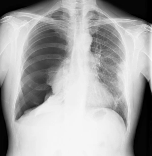
Tension pneumothorax. X-ray of the chest of a patient with tension pneumothorax that has lead to soft-tissue emphysema. Tension pneumothorax is a progressive deterioration

Background imageRespiratory Collection: English oak leaf pores, SEM

English oak leaf pores, SEM
English oak leaf pores. Coloured scanning electron micrograph (SEM) of stomata (round) on the underside of a leaf from an English oak (Quercus robur) tree

Background imageRespiratory Collection: Cystic fibrosis

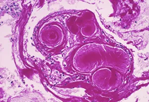
Cystic fibrosis. Light micrograph of a section through a bronchus (airway) of the lungs in a case of cystic fibrosis (mucoviscidosis)

Background imageRespiratory Collection: Paramyxovirus particles, TEM

Paramyxovirus particles, TEM
Sendai virus. Coloured transmission electron micrograph (TEM) of Sendai virus particles (virions, orange). The protein coat (capsid) of one of the particles has split

Background imageRespiratory Collection: French lavender leaf pore, SEM

French lavender leaf pore, SEM
French lavender leaf pore. Coloured scanning electron micrograph (SEM) of an open stoma (centre, black). Stomata are pores that open and close in order to regulate gas exchange in a plant

Background imageRespiratory Collection: Advertisement for ozone paper

Advertisement for ozone paper
5311927 Advertisement for ozone paper; (add.info.: Advertisement for Ozone Paper for the relief and cure of Asthma, Chronic Bronchitis and Bronchitic Asthma)

Background imageRespiratory Collection: Male chest anatomy, artwork

Male chest anatomy, artwork
Male chest anatomy, computer artwork

Background imageRespiratory Collection: 13132609

13132609
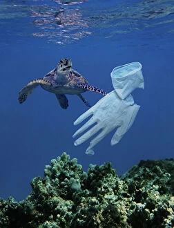
Turtle approaching surgical glove drifting in the ocean. The glove looks like a jellyfish which is one of its common foods

Background imageRespiratory Collection: 13132608

13132608
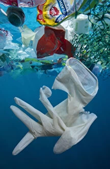
Used surgical glove drifting at sea, along with other plastic waste. Single-use masks, gloves and bottles of sanitizer shielding us from the spread of COVID-19 are ending up on the streets

Background imageRespiratory Collection: 13132607

13132607
Used surgical glove drifting at sea, along with other plastic waste. Single-use masks, gloves and bottles of sanitizer shielding us from the spread of COVID-19 are ending up on the streets

Background imageRespiratory Collection: 13132606

13132606
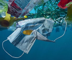
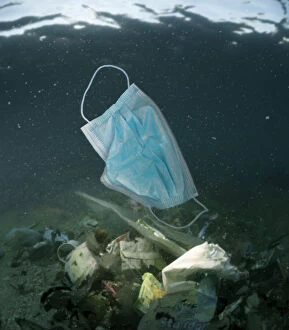
Surgical mask drifting in the ocean along with other plastic waste. Note Yellow Pygmy Goby, Lubricogobius exiguus, (about 2cm size) hiding inside the mask folds

Background imageRespiratory Collection: 13132605

13132605
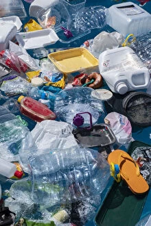
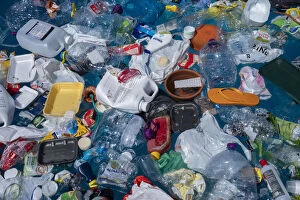
Plastic garbage floating in the ocean. The Covid-19 crisis has spurred rapid production of surgical masks, gloves, and other protective equipment

Background imageRespiratory Collection: 13132604

13132604
Plastic garbage floating in the ocean. Single-use masks, gloves and bottles of sanitizer shielding us from the spread of COVID-19 are ending up on the streets, in the seas and among wildlife

Background imageRespiratory Collection: 13132603

13132603
Used surgical mask used adrift at sea, along with other plastic waste. Single-use masks, gloves and bottles of sanitizer shielding us from the spread of COVID-19 are ending up on the streets

Background imageRespiratory Collection: 13132602

13132602
Concept image depicting the ocean pollution by plastic gloves and masks related with coronavirus disease. The Covid-19 crisis has spurred rapid production of surgical masks, gloves

Background imageRespiratory Collection: 13132601

13132601
Hawksbill Turtle approaching surgical glove drifting in the ocean. The glove looks like a jellyfish which is one of its common foods.Single-use masks

Background imageRespiratory Collection: 13132600

13132600
Turtle approaching surgical glove drifting in the ocean. The glove looks like a jellyfish which is one of its common foods

Background imageRespiratory Collection: 13132599

13132599
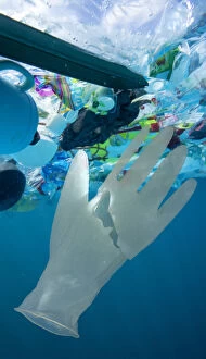
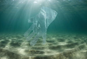
Transparent plastic glove drifting in the ocean. Single-use masks, gloves and bottles of sanitizer shielding us from the spread of COVID-19 are ending up on the streets

Background imageRespiratory Collection: 13132598

13132598
Used masks and surgical gloves at the waters edge in an estuary. Single-use masks, gloves and bottles of sanitizer shielding us from the spread of COVID-19 are ending up on the streets

Background imageRespiratory Collection: 13132597

13132597
Used masks and surgical gloves at the waters edge in an estuary. The Covid-19 crisis has spurred rapid production of surgical masks, gloves, and other protective equipment

Background imageRespiratory Collection: 13132596

13132596
Mask and surgical gloves on top of urban sewer grid. Single-use masks, gloves and bottles of sanitizer shielding us from the spread of COVID-19 are ending up on the streets

Background imageRespiratory Collection: 13132595

13132595
Mask and surgical gloves on top of urban sewer grid. Single-use masks, gloves and bottles of sanitizer shielding us from the spread of COVID-19 are ending up on the streets

Background imageRespiratory Collection: Human respiratory organs (colour litho)

Human respiratory organs (colour litho)
3097109 Human respiratory organs (colour litho) by German School, (19th century); Private Collection; (add.info.: Human respiratory organs)

Background imageRespiratory Collection: The Respirator for colds, cough, sore throat, etc

The Respirator for colds, cough, sore throat, etc
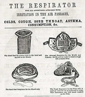
The Respirator for all affections attended with irritation in the air passages, as colds, cough, sore throat, asthma, consumption, etc. 1858

Background imageRespiratory Collection: Advertisement for Geraudels Pastilles

Advertisement for Geraudels Pastilles. 1893

Background imageRespiratory Collection: Circulatory system of heart and lungs

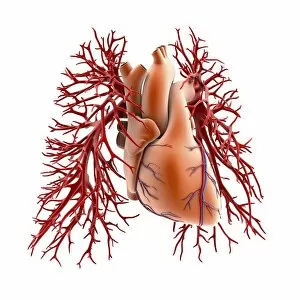
Circulatory system of heart and lungs, computer artwork. The heart (centre) is a hollow muscle that pumps blood around the body

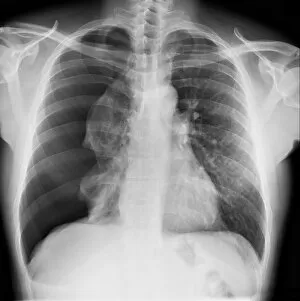
Background imageRespiratory Collection: Pneumothorax, X-ray C017 / 7148

Pneumothorax, X-ray C017 / 7148
Pneumothorax. X-ray of the chest of a 50 year old male patient with a pneumothorax, or collapsed lung. The right lung (left on X-ray) has collapsed due to a build-up of air (solid grey)

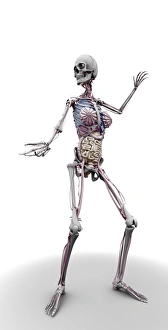
Background imageRespiratory Collection: Female skeleton and organs, artwork

Female skeleton and organs, artwork
Female skeleton and organs and blood vessels, artwork

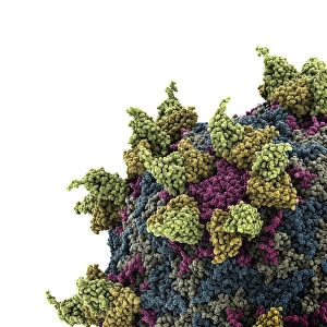
Background imageRespiratory Collection: Rhinovirus and antibody, molecular model C015 / 7139

Rhinovirus and antibody, molecular model C015 / 7139
Rhinovirus. Molecular model of the antigen-binding fragment (Fab) from a strongly neutralising antibody bound to a human rhinovirus 14 (HRV-14) particle

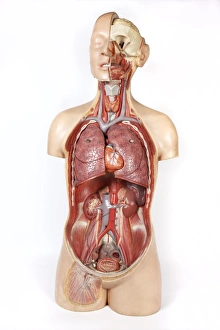
Background imageRespiratory Collection: Female anatomy, historical model

Female anatomy, historical model
Female anatomy. 1920s anatomical model showing the internal organs of the female upper body with the intestines, stomach and liver removed

Background imageRespiratory Collection: Advert for Dickins - Jones corsets 1909

Advert for Dickins - Jones corsets 1909
The Specialite Guninea corset, available from Dickins & Jones, is ideal for slight figures. It promises not to cause undue pressure to the respiratory organs. 1909

Background imageRespiratory Collection: Trachea lining, SEM C015 / 9936

Trachea lining, SEM C015 / 9936
Trachea lining. Coloured scanning electron micrograph (SEM) of a section through the lining of the trachea (wind pipe). The trachea links the larynx (voice box) to the lungs

Background imageRespiratory Collection: Rhinovirus and antibody, molecular model C015 / 7138

Rhinovirus and antibody, molecular model C015 / 7138
Rhinovirus. Molecular model of the antigen-binding fragment (Fab) from a strongly neutralising antibody bound to a human rhinovirus 14 (HRV-14) particle

Background imageRespiratory Collection: Heart and lung disease, X-ray C018 / 0498

Heart and lung disease, X-ray C018 / 0498
Heart and lung disease. Coloured X-ray of the chest of an 80 year old patient with heart and lung disease. The patient is a former miner and has chronic obstructive pulmonary disease (COPD)

Background imageRespiratory Collection: Influenza virus particles, TEM

Influenza virus particles, TEM
Influenza virus particles, coloured transmission electron micrograph (TEM). The virus consists of ribonucleic acid (RNA), surrounded by a nucleocapsid (blue) and a lipid envelope (green)

Background imageRespiratory Collection: Potato Leaf Stomata (SEM)

Potato Leaf Stomata (SEM)
Potato Stomata. Scanning electron micrographs (SEM) of open stomata on a potato leaf (Solanum tuberosum). Stomata are pores that open and close in order to regulate gas exchange in a plant

Background imageRespiratory Collection: Pneumothorax, X-ray C017 / 7812

Pneumothorax, X-ray C017 / 7812
Pneumothorax. X-ray of the chest of a patient with a pneumothorax, or collapsed lung. The right lung (left on X-ray) has collapsed due to a build-up of air (solid grey)

Background imageRespiratory Collection: LCC London Fire Brigade asbestos protective suit

LCC London Fire Brigade asbestos protective suit
Carried on emergency tenders (ETs) an asbestos suit allowed a firefighter to approach a serious fire at close quarters for the purpose of closing down a control valve or a similar task

Background imageRespiratory Collection: Allergens in trachea

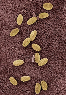
Allergens in trachea. Coloured scanning electron micrograph (SEM) of the surface of the trachea (windpipe) with a number of pollen grains (yellow) attached

Background imageRespiratory Collection: Cow anatomy, artwork

Cow anatomy, artwork
Cow anatomy. Computer artwork showing the internal anatomy of a domesticated cow (Bos primigenius). Anatomical features shown here include the skeleton, lungs, intestines, bladder

Background imageRespiratory Collection: Infectious bronchitis virus (IBV), TEM

Infectious bronchitis virus (IBV), TEM
Infectious bronchitis virus (IBV) particles, coloured transmission electron micrograph (TEM). This virus is a member of the coronavirus family

Background imageRespiratory Collection: German gas masks

German gas masks
A British impression of the type of respiratory equipment used by German soldiers during gas attacks upon Allied troops. The Germans first used poisonous gas at Ypres on April 22nd 1915

Background imageRespiratory Collection: Trade card for Dr Zed syrup, girl with pug dog

Trade card for Dr Zed syrup, girl with pug dog
Trade card for Dr Zed syrup, showing a little girl in a pink dress hugging her pug dog. Date: circa 1900s


All Professionally Made to Order for Quick Shipping

"The Marvels of the Respiratory System: Exploring the Gateway to Life" Diagram of the Lungs and Bronchial Tubes: Unveiling the intricate network within our bodies that facilitates every breath we take, this diagram showcases the lungs and bronchial tubes, highlighting their vital role in oxygenating our bloodstream. Diagram of the Heart, Lungs, and Windpipe: Witnessing a symphony of life-sustaining organs working harmoniously together, this diagram depicts the heart pumping blood while being surrounded by the lungs and windpipe – a testament to our body's remarkable respiratory system. Illustration of Human Respiratory System: Embark on an anatomical journey through our respiratory system as this illustration reveals its various components such as oral cavity, nasal cavity, larynx, trachea, bronchus, and lungs – each playing a crucial part in ensuring efficient respiration. Tension Pneumothorax X-ray: Peering into medical imaging technology's window into diagnosis and treatment strategies for collapsed lungs caused by tension pneumothorax - an urgent condition requiring immediate attention to restore normal breathing patterns. English Oak Leaf Pores SEM Image: Delving into nature's wonders at microscopic levels reveals mesmerizing images showcasing English oak leaf pores under scanning electron microscopy (SEM), reminding us how even plants have their unique respiratory systems. Cystic Fibrosis: Unmasking a Genetic Challenge Exploring cystic fibrosis - a genetic disorder affecting lung function - sheds light on ongoing research efforts aimed at improving treatments for individuals facing challenges with their respiratory health from an early age. Konstantin Buteyko: Soviet Doctor Revolutionizing Breathing Techniques Discovering how Dr. Konstantin Buteyko revolutionized breathing techniques offers insights into his innovative methods that have helped countless individuals manage asthma symptoms and enhance overall respiratory well-being. Paramyxovirus Particles TEM Image.