
Retinal Collection
"Exploring the Intricacies of Retinal: Unveiling the Wonders of Vision" The retina, also known as the innermost layer of the eye


All Professionally Made to Order for Quick Shipping
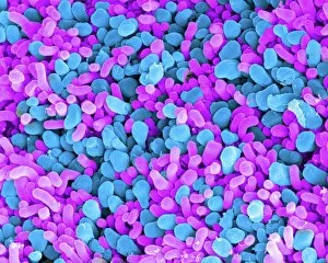
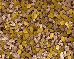
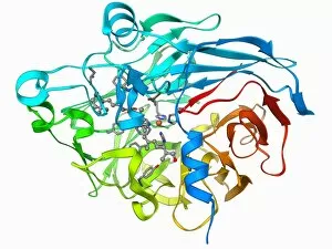
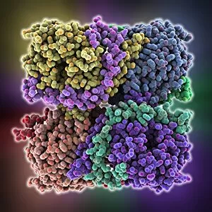
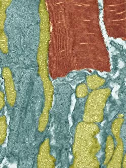
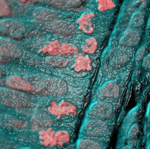
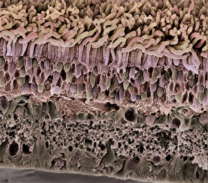
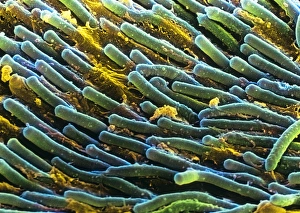
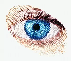
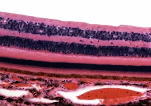
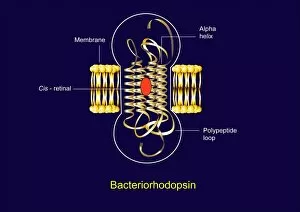
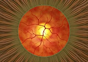
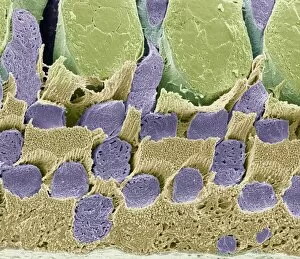
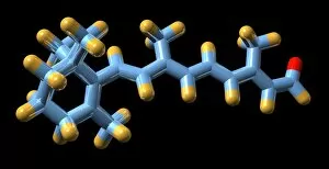
"Exploring the Intricacies of Retinal: Unveiling the Wonders of Vision" The retina, also known as the innermost layer of the eye, plays a vital role in our visual perception. Composed of specialized cells called rod and cone cells, it acts as a gateway for light to be transformed into electrical signals that are then transmitted to the brain. Through scanning electron microscopy (SEM), we can observe the intricate structure of these rod and cone cells (C014 / 4866). These microscopic wonders are responsible for different aspects of vision - rods excel in low-light conditions while cones enable color perception. Another fascinating aspect captured by SEM is the network of blood vessels within the retina. These delicate vessels ensure proper nourishment and oxygen supply to this essential sensory tissue (Retina blood vessels, SEM). Zooming further into retinal exploration, transmission electron microscopy (TEM) reveals intriguing details such as Rift Valley fever virus particles present within retinal tissues. This highlights how viruses can impact ocular health on a microscopic level (Rift Valley fever virus, TEM). Delving deeper into molecular structures involved in vision, we encounter rhodopsin protein molecules. Rhodopsin is an integral component found in rod cells that enables them to detect light stimuli efficiently (Rhodopsin protein molecule). Its interaction with light triggers a cascade leading to signal transduction processes crucial for sight. Microscopic examination using advanced techniques like TEM allows us to witness mesmerizing images showcasing various components involved in visual processing. From bacteriorhodopsin proteins associated with photosynthesis-like reactions (Bacteriorhodopsin protein) to metarhodopsin molecules contributing to phototransduction pathways (F006 / 9709), each element unravels new insights about our remarkable sense of sight.