
Sagittal Collection
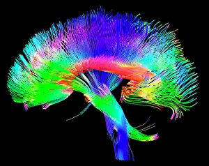
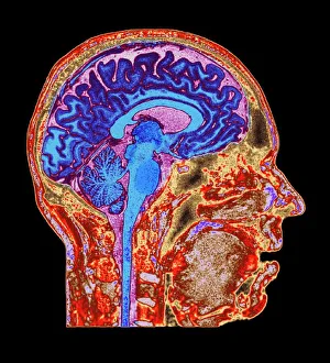
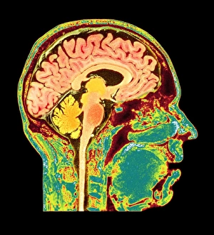
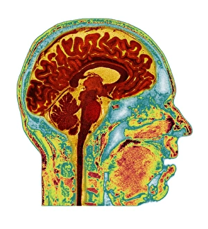
"Sagittal: Unlocking the Mysteries of the Human Brain and Body" Delve into the intricate web of brain pathways as we explore the fascinating world anatomy


All Professionally Made to Order for Quick Shipping
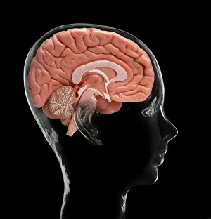
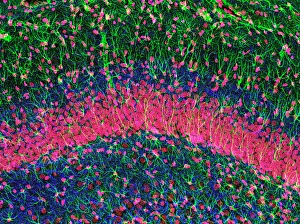
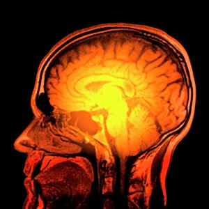
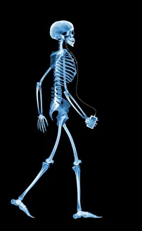
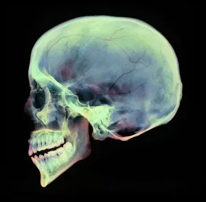
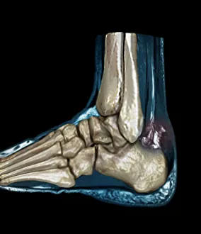
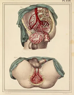
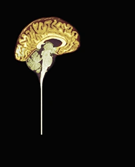
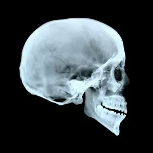
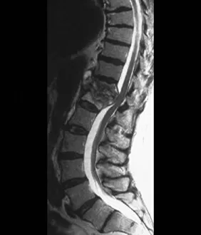
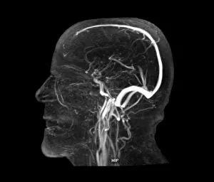
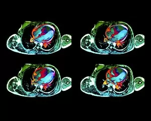
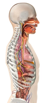
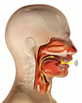
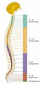
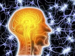
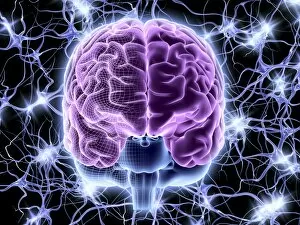
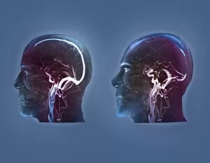
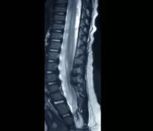
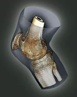
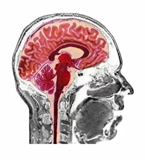
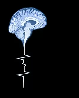
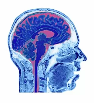
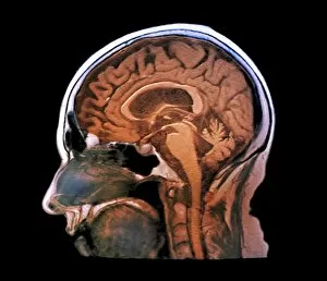
"Sagittal: Unlocking the Mysteries of the Human Brain and Body" Delve into the intricate web of brain pathways as we explore the fascinating world anatomy. From studying the human brain model to examining hippocampus brain tissue, this captivating field offers a glimpse into our most complex organ. Peering through an MRI scan, witness the mesmerizing intricacies of brain anatomy unfold before your eyes. Discover how these scans provide invaluable insights into neurological conditions and aid in medical diagnoses. But it's not just the brain that holds secrets; even skeletons have stories to tell. Observe a skeleton drinking on an X-ray image, reminding us that every part of our body is interconnected. Moving beyond bones, delve deeper into medical artistry with X-rays revealing hidden details within a human skull. These images showcase both beauty and fragility, highlighting our vulnerability as well as resilience. Travel back in time with an 1825 artwork depicting male groin arteries—a testament to how far medical knowledge has progressed over centuries. Witness how science and art intertwine to unravel mysteries once shrouded in uncertainty. Shift focus from historical illustrations to modern-day medicine as we examine an MRI scan capturing a ruptured Achilles tendon. This visual representation underscores both injury and healing processes within our bodies. Returning to normalcy, marvel at pristine MRI scans showcasing healthy human brains—reminders of what lies beneath each individual's unique thoughts and emotions. Appreciate their complexity while acknowledging their fragility. Zoom out from singular organs to encompass both brain and spinal cord in one comprehensive MRI image—an awe-inspiring reminder of their interdependence for proper bodily function. Concluding this journey through sagittal exploration is an artistic rendition of a brain scan—a fusion between science and creativity that captures its essence beyond mere scientific documentation. Sagittal reveals more than meets the eye—it unravels connections between mind, body, history, artistry, injury, healthiness—all encapsulated within a single word.