Fraud Blocker Skip to main content

Sectioned Collection

"Exploring the Intricacies: From Cerebellum Tissue to Fusion Research, Unveiling the World Sectioned" Delving into the depths of scientific exploration

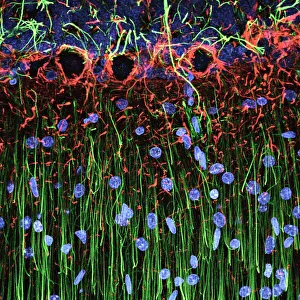
Background imageSectioned Collection: Cerebellum tissue, light micrograph

Cerebellum tissue, light micrograph
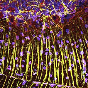
Cerebellum tissue. Confocal light micrograph of a section through the cerebellum of the brain. Purkinje cells, a type of neuron (nerve cell), are red

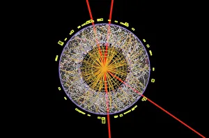
Background imageSectioned Collection: Higgs boson event, ATLAS detector C013 / 6892

Higgs boson event, ATLAS detector C013 / 6892
Higgs boson event. Graphic of a transverse section through a detector showing one of the numerous particle collision events recorded during the search for the Higgs boson

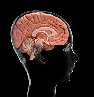
Background imageSectioned Collection: Human brain, model

Human brain, model. Composite image of a sectioned model human brain inside a glass head. This view shows some of the brains major structures, including: the cerebral cortex (folded areas of tissue)

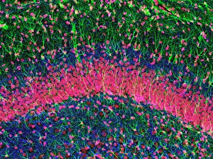
Background imageSectioned Collection: Hippocampus brain tissue

Hippocampus brain tissue
Hippocampus tissue. Light micrograph of a sagittal (side view) section through the hippocampus of the brain showing the nerve cells within it

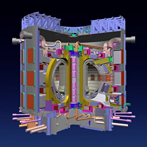
Background imageSectioned Collection: Fusion research, tokamak device

Fusion research, tokamak device
Nuclear fusion research. Cutaway computer artwork of the planned ITER tokamak device. It will be the first full scale fusion device to produce energy at a level of an electricity-producing power

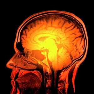
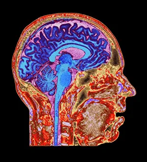
Background imageSectioned Collection: Brain anatomy, MRI scan

Brain anatomy, MRI scan
Brain anatomy. Coloured magnetic resonance imaging (MRI) scan of the human head from the side. The sagittal scan has halved the brain, revealing the internal anatomy

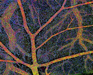
Background imageSectioned Collection: Brain tissue blood supply

Brain tissue blood supply. Light micrograph of a section through cortex tissue from a brain, showing the blood vessels (branching) that supply it

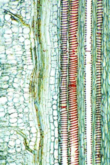
Background imageSectioned Collection: Dicotyledon plant stem, light micrograph

Dicotyledon plant stem, light micrograph
Dicotyledon plant stem. Light micrograph of a longitudinal radial section through a typical dicotyledon stem. The section has passed through a vascular bundle and other stem tissue

Background imageSectioned Collection: EDTA crystals, light micrograph

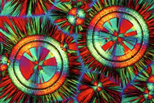
EDTA crystals, light micrograph
EDTA crystals. Polarised light micrograph of a section through ethylenediaminetetraacetic acid (EDTA) crystals. EDTA is used primarily for its effectiveness at binding metal ions (chelating)

Background imageSectioned Collection: Botanik Digitalis purpurea L. Fingerhut 160: 1

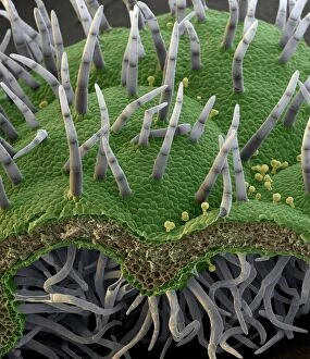
Botanik Digitalis purpurea L. Fingerhut 160: 1
Foxglove leaf. Coloured scanning electron micrograph (SEM) of a section through a leaf from a foxglove (Digitalis purpurea) plant, showing its hairs (trichomes)

Background imageSectioned Collection: Kidney tubules in section

Kidney tubules in section
Kidney tubules. Fluorescent light micrograph of a section through kidney tissue showing numerous tubules (black/green). The tubules are seen in cross-section
Background imageSectioned Collection: Optical computing

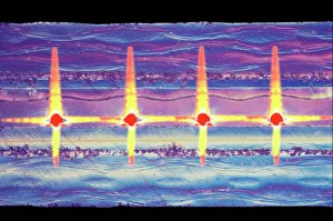
Optical computing. Coloured light micrograph of a section through a hybrid electronic-optical circuit board. Optical waveguides (red) are used to transmit data in the form of light pulses

Background imageSectioned Collection: Human skull, X-ray

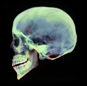
Human skull, X-ray
Human skull. Coloured X-ray of a sagittal section through a human skull. The skull has been sliced in half down the centre, revealing details of the internal structures

Background imageSectioned Collection: Rough endoplasmic reticulum, TEM

Rough endoplasmic reticulum, TEM
Rough endoplasmic reticulum, coloured transmission electron micrograph (TEM). This section shows the rough endoplasmic reticulum (ER, folds, centre), a membranous structure that occurs in cells

Background imageSectioned Collection: Alzheimers brain, computer artwork

Alzheimers brain, computer artwork
Alzheimers brain. Computer graphic of a vertical (coronal) slice through the brain of an Alzheimer patient (at left) compared with a normal brain (at right)

Background imageSectioned Collection: Eye anatomy, artwork

Eye anatomy, artwork
Eye anatomy, computer artwork. At the front of the eye is the cornea, a transparent coating. Behind this is the lens, which is partly covered by the iris

Background imageSectioned Collection: Castor oil stem, light micrograph

Castor oil stem, light micrograph
Castor oil stem. Light micrograph of a longitudinal section through the stem of a castor oil (Ricinus communis) plant. At right are large and small parenchyma cells (blue)

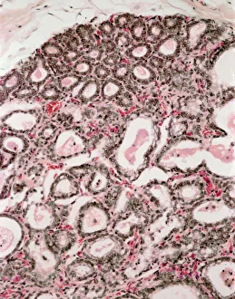
Background imageSectioned Collection: Lactating breast tissue, light micrograph

Lactating breast tissue, light micrograph
Lactating breast tissue. Light micrograph of a section through healthy breast (mammary gland) tissue during lactation. Glandular tissue in the breast has proliferated to produce spaces (alveoli)

Background imageSectioned Collection: Alzheimers brain

Alzheimers brain. Computer processed vertical (coronal) slice through the brain of an Alzheimer patient (at left) compared with a normal brain (at right)

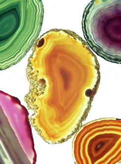
Background imageSectioned Collection: Cut agates

Cut agates. Assortment of cut and polished agates. Agate is a variety of quartz (silicon oxide). The colours are produced by impurities in the mineral
Background imageSectioned Collection: Pine pollen grains, light micrograph

Pine pollen grains, light micrograph
Pine pollen grains. Light micrograph of a section through winged pollen grains (microspores) from a pine (Pinus sp.) tree

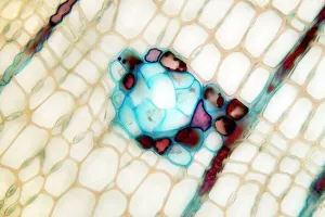
Background imageSectioned Collection: Pine stem, light micrograph

Pine stem, light micrograph
Pine stem. Light micrograph of a section through the stem of a pine (Pinus sp.) tree, showing xylem tissue. The xylem is made up of tracheid cells (light pink)

Background imageSectioned Collection: Lime tree stem, light micrograph

Lime tree stem, light micrograph
Lime tree stem. Light micrograph of a section through the stem of a lime tree (Tilia europaea). The outer epidermis has been shed and replaced by a layer of cork (dark red)

Background imageSectioned Collection: Cell types, artwork

Cell types, artwork
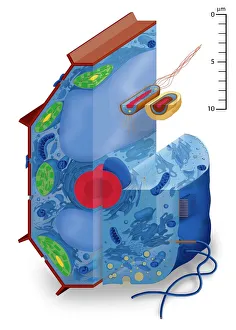
Cell types. Cutaway artwork with a scale bar (upper right, in micrometres) showing the relative sizes of eukaryotic cells (those containing a nucleus) and prokaryotic cells (those lacking a nucleus)

Background imageSectioned Collection: Compact bone, light micrograph

Compact bone, light micrograph
Compact bone. Polarised light micrograph of a transverse section through compact bone tissue, showing Haversian canals (circular regions)

Background imageSectioned Collection: Fat cells, TEM

Fat cells, TEM
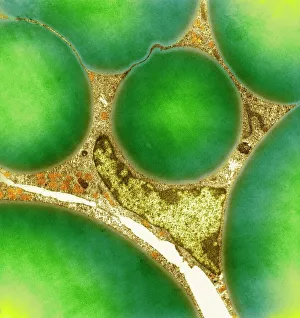
Fat cells. Coloured transmission electron micrograph (TEM) of a section through fat (adipose) tissue, consisting of fat-storing cells (adipocytes, green) supported by connective tissue

Background imageSectioned Collection: Jelly fish, artwork

Jelly fish, artwork
Jelly fish (Leonura terminalis). Historical artwork of a deep sea jelly fish observed by Ernst Haeckel during an expedition on HMS Challenger (1873-1876)

Background imageSectioned Collection: Cerebellum tissue, light micrograph

Cerebellum tissue, light micrograph
Cerebellum tissue. Confocal light micrograph of a section through the cerebellum of the brain showing two types of glial cells (support cells); astrocytes (star-shaped)

Background imageSectioned Collection: Brain motor cortex pathways, artwork C016 / 6532

Brain motor cortex pathways, artwork C016 / 6532
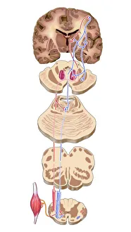
Brain motor cortex pathways. Artwork of a sectioned human brain, brainstem and spinal cord, showing neural pathways (red and blue) from the motor cortex of the brain

Background imageSectioned Collection: Bacterial meningitis, MRI scan

Bacterial meningitis, MRI scan
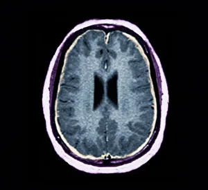
Bacterial meningitis. Coloured magnetic resonance imaging (MRI) scan of an axial section through the brain of a 16-year-old patient

Background imageSectioned Collection: Skull anatomy

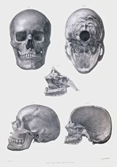
Skull anatomy. Historical anatomical artwork of various views of the human skull. The frontal view (upper left) shows the teeth, nose and eye spaces

Background imageSectioned Collection: Tendon, SEM

Tendon, SEM
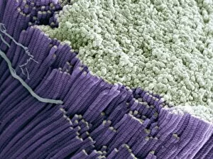
Tendon, coloured scanning electron micrograph (SEM), showing bundles of collagen fibres. Magnification: x5000 when printed at 10 centimetres wide

Background imageSectioned Collection: Maize root, light micrograph

Maize root, light micrograph
Maize root. Light micrograph of a section through the root of a maize plant (Zea mays) showing a vascular cylinder (centre)

Background imageSectioned Collection: Water lily leaf, light micrograph

Water lily leaf, light micrograph
Water lily leaf. Light micrograph of a transverse section through the leaf of a water lily (Nympha sp.) plant. All aquatic plants (hydrophytes) have a similar structure

Background imageSectioned Collection: Cerebellum tissue, light micrograph

Cerebellum tissue, light micrograph
Cerebellum tissue. Confocal light micrograph of a section through the cerebellum of the brain. Purkinje cells, a type of neuron (nerve cell), are red

Background imageSectioned Collection: Normal human brain, MRI scan C016 / 8845

Normal human brain, MRI scan C016 / 8845
Brain. Coloured magnetic resonance imaging (MRI) scan of a sagittal section through a patients head showing a healthy human brain

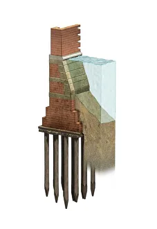
Background imageSectioned Collection: Venice building foundations, artwork C016 / 7703

Venice building foundations, artwork C016 / 7703
Venice building foundations. Computer artwork showing the foundations of a typical building in Venice, Italy. Venice is built on the marshy Venetian Lagoon which stretches along the shoreline

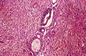
Background imageSectioned Collection: Ovarian cancer, light micrograph C015 / 7103

Ovarian cancer, light micrograph C015 / 7103
Ovary cancer. Light micrograph of a section through a Krukenberg tumour (down centre) of the ovary. This is a secondary cancer that spreads (metastasises) from the digestive system

Background imageSectioned Collection: Liver tissue cirrhosis, light micrograph

Liver tissue cirrhosis, light micrograph
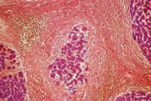
Liver cirrhosis. Light micrograph of a section through liver tissue showing cirrhosis. This is a condition where the liver responds to the injury or death of some of its cells by producing

Background imageSectioned Collection: Cystic fibrosis

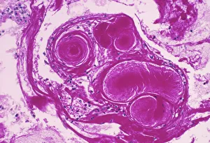
Cystic fibrosis. Light micrograph of a section through a bronchus (airway) of the lungs in a case of cystic fibrosis (mucoviscidosis)

Background imageSectioned Collection: Ammonite

Ammonite. Polished sectioned ammonite fossil. Ammonites are extinct marine invertebrates. They first appeared in the Late Silurian to Early Devonian period (around 400 million years ago)

Background imageSectioned Collection: Lung tissue, TEM

Lung tissue, TEM
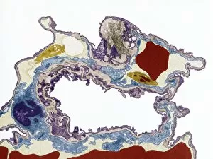
Lung tissue, coloured transmission electron micrograph (TEM). A single alveolus is at center. At top right is a pulmonary capillary containing a red blood cell (red) and a platelet (yellow)

Background imageSectioned Collection: Islet of Langerhans, light micrograph

Islet of Langerhans, light micrograph
Islet of Langerhans. Light micrograph of a section through an islet of Langerhans (centre) in the pancreas. This structure is composed of clumps of secretory cells

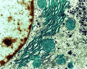
Background imageSectioned Collection: Golgi apparatus, SEM

Golgi apparatus, SEM
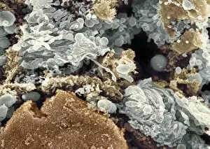
Golgi apparatus, coloured scanning electron micrograph (SEM). Section through a liver cell showing its Golgi apparatus (grey), a membrane-bound organelle that modifies and packages proteins

Background imageSectioned Collection: Cotton plant leaf, light micrograph

Cotton plant leaf, light micrograph
Cotton plant leaf. Light micrograph of a transverse section through a leaf of the cotton plant (Gossypium hirsutum). Magnification: x45 when printed at 10 centimetres wide

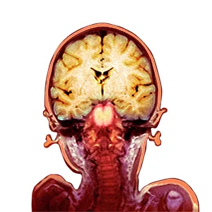
Background imageSectioned Collection: Childs brain, MRI scan

Childs brain, MRI scan
Childs brain. Coloured magnetic resonance imaging (MRI) scan of an coronal (vertical) section through the head and brain of a healthy 3-year-old child, passing through the ears

Background imageSectioned Collection: Silver birch twig, SEM

Silver birch twig, SEM
Silver birch twig. Coloured scanning electron micrograph (SEM) of a section through a silver birch (Betula pendula) twig. At the centre of the twig (top centre left) is the pith

Background imageSectioned Collection: Collagen fibres

Collagen fibres. Coloured transmission electron micrograph (TEM) of a section through healthy collagen fibres from human skin


All Professionally Made to Order for Quick Shipping

"Exploring the Intricacies: From Cerebellum Tissue to Fusion Research, Unveiling the World Sectioned" Delving into the depths of scientific exploration, a light micrograph reveals the intricate structure of cerebellum tissue. Witnessing a Higgs boson event through the lens of ATLAS detector C013/6892, scientists capture an awe-inspiring moment in particle physics. A meticulously crafted model showcases the complexity and beauty of the human brain, inviting us to marvel at its wonders. Peering into hippocampus brain tissue under a microscope, we uncover secrets that may hold keys to memory formation and learning processes. In pursuit of limitless energy sources, fusion research takes center stage as we examine a tokamak device designed for controlled nuclear fusion reactions. The inner workings of our brains come alive through an MRI scan, revealing detailed brain anatomy like never before imagined. Illuminated by light microscopy, dicotyledon plant stem sections unveil nature's architectural masterpiece in stunning detail. EDTA crystals take on mesmerizing patterns when viewed under intense magnification - showcasing their unique molecular arrangement through light micrography. Journeying deep within our bodies, kidney tubules are sectioned to shed light on their vital role in maintaining fluid balance and waste removal systems. An X-ray image exposes every contour and intricacy of a human skull – offering glimpses into our skeletal framework with remarkable clarity. Through transmission electron microscopy (TEM), rough endoplasmic reticulum emerges as an interconnected network responsible for protein synthesis within cells' cytoplasmic matrix Computer artwork visualizes Alzheimer's-affected brain regions – reminding us why continued research is crucial in unraveling this devastating disease. In this captivating journey across various scientific disciplines and microscopic landscapes, "sectioned" unveils hidden worlds that expand our understanding of the universe and ourselves.