
Single Celled Collection
"Unveiling the Microscopic World: Captivating Single-Celled Organisms Under the SEM" Delicate and intricate


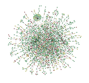


All Professionally Made to Order for Quick Shipping
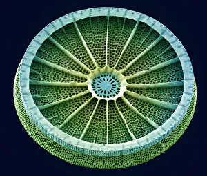
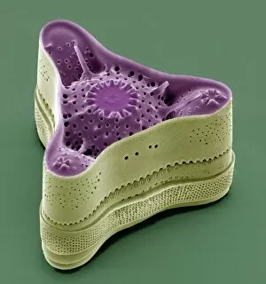
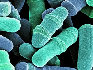
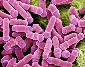
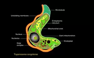
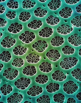
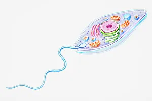
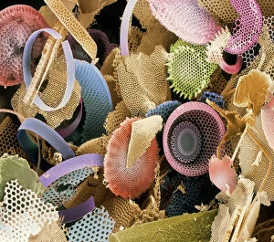
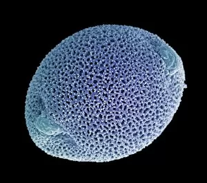
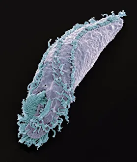
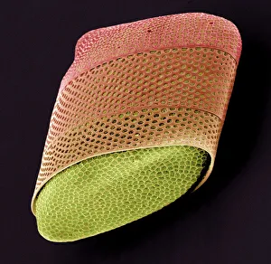
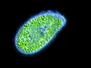
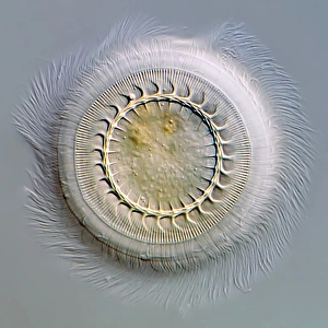
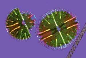
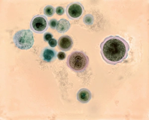
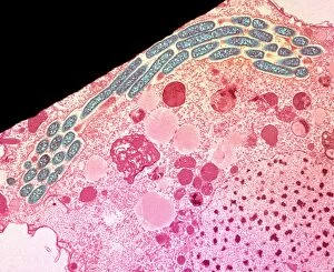
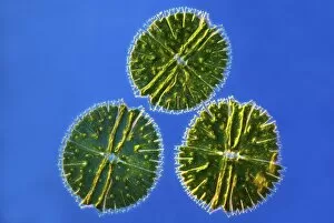
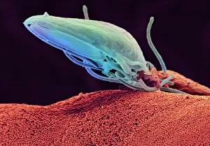
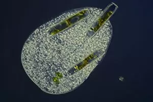
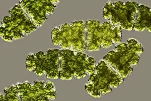
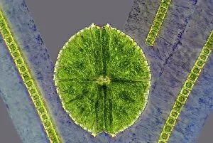
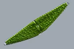
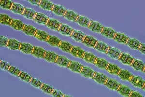
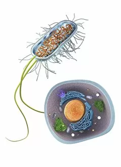
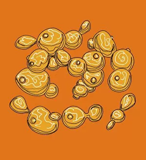
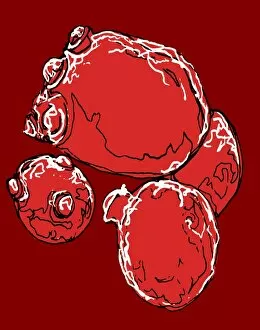
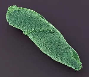
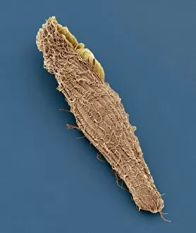
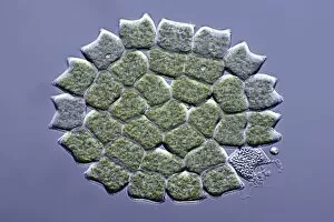
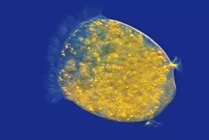
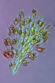
"Unveiling the Microscopic World: Captivating Single-Celled Organisms Under the SEM" Delicate and intricate, diatoms reveal their mesmerizing beauty under the scanning electron microscope (SEM). Their symmetrical frustules showcase nature's artistry at its finest. Witness the fascinating process of cell division in yeast cells as they multiply and propagate. The SEM captures this microscopic dance with remarkable detail. Behold the captivating artwork of Trypanosome protozoan, a single-celled organism that evokes both awe and curiosity. Its unique form is brought to life through artistic interpretation. Diatom alga, another marvel of nature, showcases its stunning structure when examined under the SEM lens. These tiny organisms play a crucial role in aquatic ecosystems worldwide. Protozoa, scavengers extraordinaire. Discover these single-celled wonders as they scavenge for particles and microorganisms or absorb nutrients from their environment – truly masters of survival. Acrosphaera radiolarian takes center stage with its intricate skeletal structure captured by SEM imaging techniques. Marvel at this ancient marine creature frozen in time. Journey back millions of years as fossilized diatoms come into focus under the SEM lens, offering glimpses into Earth's past and providing valuable insights for scientific research today. Diatom frustule - an architectural masterpiece on a microscopic scale. Explore these silica-based shells that protect diatoms while showcasing an array of breathtaking patterns through SEM imagery. Oxytricha ciliate protozoan reveals its astonishing complexity when observed using advanced microscopy techniques like SEM C019 / 0253 – unlocking secrets hidden within this enigmatic world. Exploring single-celled organisms through scanning electron microscopy opens up a whole new dimension where beauty meets science; revealing intricacies unseen by our naked eyes but essential to understanding life's diversity on our planet.