
Sinus Collection
"Exploring the Intricacies of Sinus: Unveiling the Hidden Depths" Delving into the depths of human anatomy


All Professionally Made to Order for Quick Shipping
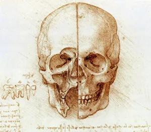
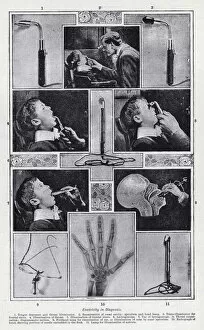
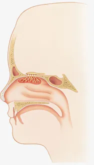
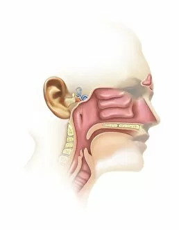
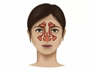
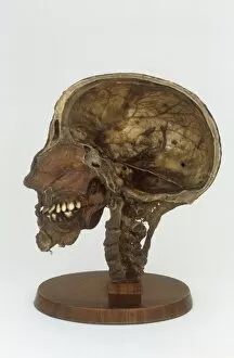
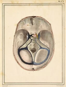
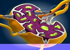
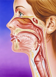
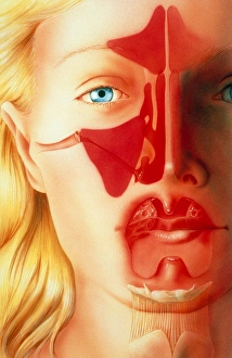
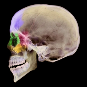
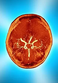
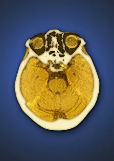
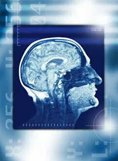
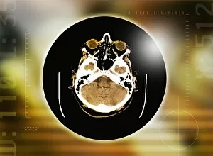
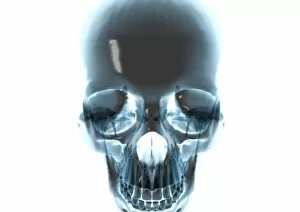
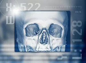
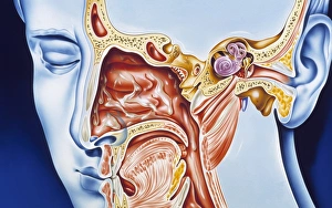
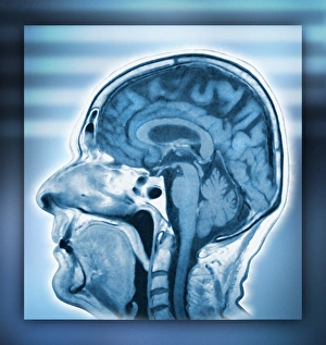
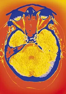
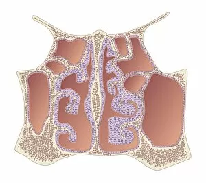
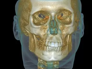
"Exploring the Intricacies of Sinus: Unveiling the Hidden Depths" Delving into the depths of human anatomy, Leonardo da Vinci's intricate sketches reveal the complexity of skull anatomy, shedding light on the enigmatic sinuses. A CT scan captures a detailed image of nose and sinuses, providing a window into these vital structures that often go unnoticed in our daily lives. The marble portrait bust of Severus Alexander transports us back to ancient times, reminding us that even centuries ago, people were aware of the significance health. An engraving showcasing the inner surface of occipital bone highlights its connection to sinuses and their role in maintaining overall well-being. In a black and white photo from history, electricity is utilized for diagnosis purposes, demonstrating how science has evolved in understanding sinus-related ailments over time. Through an illustration depicting smell sensors and nasal epithelium, we gain insight into how our sense of smell relies on various components such as olfactory bulbs and turbinate bones within our nasal cavity. Exploring further into human anatomy reveals not only intricate details about inner ear but also uncovers connections between this delicate system and our sinuses. A microscopic close-up unveils the common cold influenza virus lurking within our bodies – a reminder that sinus health plays a crucial role in warding off such infections. Detailed illustrations guide us through an exploration of nasal sinuses' complex structure, emphasizing their importance in maintaining respiratory function and overall well-being. Peering inside with fascination at an illustration displaying dura mater sinuses atop a human brain offers insights into how these interconnected systems work together seamlessly for optimal functioning. From another perspective comes an illustration showcasing side view dura mater sinuses alongside a human brain – highlighting their integral role in cerebral circulation while hinting at potential complications related to sinus disorders.