
Skeletal Collection
Skeletal wonders unraveling the mysteries of life: From Leonardo da Vinci's intricate study of skull anatomy to the muscular system on our very own heads


All Professionally Made to Order for Quick Shipping
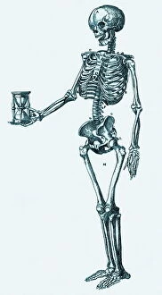
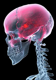
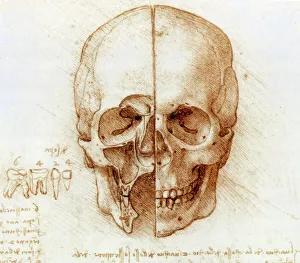
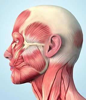
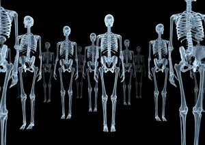
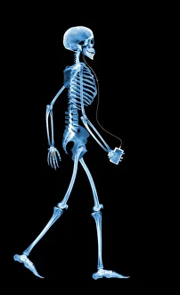
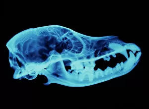
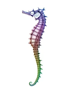
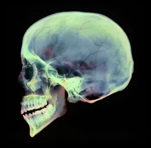
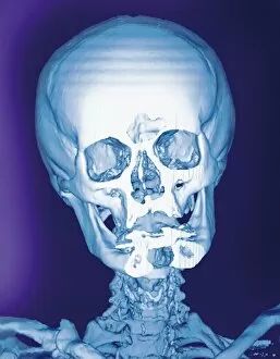
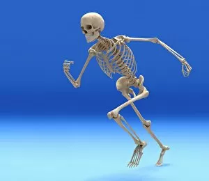
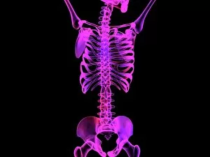
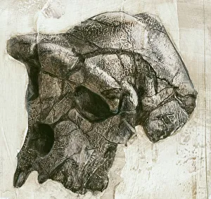
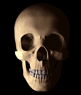
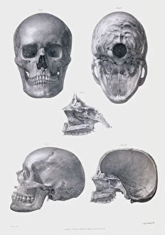
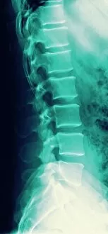
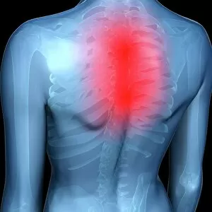
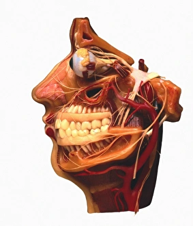
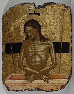
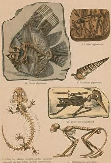
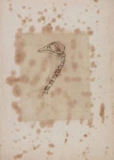
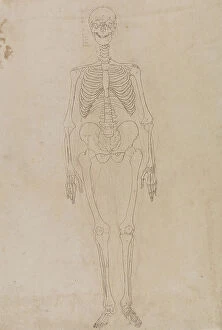
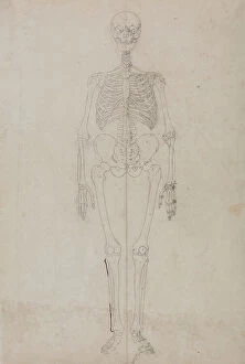
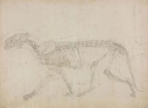
Skeletal wonders unraveling the mysteries of life: From Leonardo da Vinci's intricate study of skull anatomy to the muscular system on our very own heads, we delve into the captivating world of skeletons. Witness a skeleton indulging in a bone-chilling drink, as X-ray technology unveils its hidden secrets. Explore beyond human boundaries with an X-ray glimpse into a dog's skull and marvel at the delicate structure of a seahorse skeleton. Journey deeper into our own mortality with hauntingly beautiful X-rays capturing both human and horse skulls. Admire the artistry behind an awe-inspiring running skeleton within our bodies, portrayed through stunning artwork. Immerse yourself in ethereal X-ray masterpieces showcasing skeletal beauty from every angle. Discover Sahelanthropus tchadensis' ancient skull, bridging gaps between past and present civilizations. Finally, lose yourself in mesmerizing X-ray artwork that captures the enigmatic allure of skeletons when viewed from below. Step into this extraordinary realm where science meets art, revealing the timeless fascination held by these bony structures.