Fraud Blocker Skip to main content

Stoma Collection

Stoma, also known as leaf pores, are fascinating structures found on the surface of plant leaves

Background imageStoma Collection: Leaf pores, SEM

Leaf pores, SEM
Leaf pores. Coloured scanning electron micrograph (SEM) of stomata (holes) on the surface of a leaf. These structures perform a similar function to the pores in human skin

Background imageStoma Collection: Picture No. 11675585

Picture No. 11675585
Scanning Electron micrograph (SEM)showing stomata on a Yew Leaf. Date:

Background imageStoma Collection: English oak leaf pores, SEM

English oak leaf pores, SEM
English oak leaf pores. Coloured scanning electron micrograph (SEM) of stomata (round) on the underside of a leaf from an English oak (Quercus robur) tree

Background imageStoma Collection: Picture No. 11675628

Picture No. 11675628
Scanning Electron Micrograph (SEM): Stomata of Yew Leaf. Date:

Background imageStoma Collection: French lavender leaf pore, SEM

French lavender leaf pore, SEM
French lavender leaf pore. Coloured scanning electron micrograph (SEM) of an open stoma (centre, black). Stomata are pores that open and close in order to regulate gas exchange in a plant

Background imageStoma Collection: Illustration of cross-section through leaf, showing upper epidermis, palisade mesophyll, xylem vesse

Illustration of cross-section through leaf, showing upper epidermis, palisade mesophyll, xylem vesse

Background imageStoma Collection: Chives leaf stoma, SEM C016 / 8060

Chives leaf stoma, SEM C016 / 8060
Chives leaf stoma. Coloured scanning electron micrograph (SEM) of a stoma (oval, centre) on the surface of a leaf from a chives (Allium schoenoprasum) plant

Background imageStoma Collection: Coltsfoot leaf stomata, SEM C016 / 8052

Coltsfoot leaf stomata, SEM C016 / 8052
Coltsfoot leaf stomata. Coloured scanning electron micrograph (SEM) of two stomata (round) on the surface of a leaf from a coltsfoot (Tussilago farfara) plant

Background imageStoma Collection: Potato Leaf Stomata (SEM)

Potato Leaf Stomata (SEM)
Potato Stomata. Scanning electron micrographs (SEM) of open stomata on a potato leaf (Solanum tuberosum). Stomata are pores that open and close in order to regulate gas exchange in a plant

Background imageStoma Collection: Stomata on epidermis of Elder leaf

Stomata on epidermis of Elder leaf
Stomata. Coloured scanning electron micrograph of the under surface of a leaf from an Elder tree, Sambucus nigra, showing an open stomata

Background imageStoma Collection: Picture No. 10877020

Picture No. 10877020
Scanning Electron micrograph (SEM): Open Stoma of a plant leaf; Date:

Background imageStoma Collection: Picture No. 10877021

Picture No. 10877021
Scanning Electron micrograph (SEM): Open Stoma of a plant leaf; Date:

Background imageStoma Collection: Tobacco leaf, SEM C016 / 9465

Tobacco leaf, SEM C016 / 9465
Tobacco leaf. Coloured scanning electron micrograph (SEM) of the underside of a winged tobacco (Nicotiana alata) leaf, showing trichomes (long) and a stomata (small, oval)

Background imageStoma Collection: Squash leaf, SEM C016 / 9463

Squash leaf, SEM C016 / 9463
Squash leaf. Coloured scanning electron micrograph (SEM) of the lower surface of a squash (Curcubita maxima) leaf, showing trichomes (mushroom-shaped) and a stoma (slit, upper centre)

Background imageStoma Collection: Thale cress leaf stoma, SEM C016 / 9460

Thale cress leaf stoma, SEM C016 / 9460
Thale cress leaf stoma. Coloured scanning electron micrograph (SEM) of a stoma (round, centre) on the surface of a leaf from a thale cress (Arabidopsis thaliana) plant

Background imageStoma Collection: Thale cress leaf stoma, SEM C016 / 9459

Thale cress leaf stoma, SEM C016 / 9459
Thale cress leaf stoma. Coloured scanning electron micrograph (SEM) of a stoma (round, centre) on the surface of a leaf from a thale cress (Arabidopsis thaliana) plant

Background imageStoma Collection: Squash leaf, SEM C016 / 9464

Squash leaf, SEM C016 / 9464
Squash leaf. Coloured scanning electron micrograph (SEM) of the underside of a squash (Curcubita maxima) leaf, showing trichomes (mushroom-shaped) and a stoma (slit, upper centre)

Background imageStoma Collection: Plant stoma, ESEM

Plant stoma, ESEM
Plant stoma, coloured environmental scanning electron micrograph (ESEM). A stoma is a pore that regulates the exchange of gases and water vapour into and out of the plant

Background imageStoma Collection: Chloroplast, TEM C016 / 6297

Chloroplast, TEM C016 / 6297
Chloroplast. Coloured transmission electron micrograph (TEM) of chloroplast from the moss Physcomitrella patens. Chloroplasts are the sites of photosynthesis

Background imageStoma Collection: Chloroplast, TEM C016 / 6298

Chloroplast, TEM C016 / 6298
Chloroplast. Coloured transmission electron micrograph (TEM) of chloroplast from the moss Physcomitrella patens. Chloroplasts are the sites of photosynthesis

Background imageStoma Collection: Grape leaf stoma, SEM C014 / 4740

Grape leaf stoma, SEM C014 / 4740
Grape leaf stoma. Coloured scanning electron micrograph (SEM) of a closed stoma (centre-right) on a leaf from a grape (Vitis sp.) vine

Background imageStoma Collection: Plant water transport, artwork

Plant water transport, artwork
Plant water transport. Artwork of water molecules (blue) entering a tree through osmosis in the roots, passing upwards through the xylem vessels in the trunk and branches

Background imageStoma Collection: Sorrel leaf stoma, SEM C016 / 8059

Sorrel leaf stoma, SEM C016 / 8059
Sorrel leaf stoma. Coloured scanning electron micrograph (SEM) of a stoma (round, centre) on the surface of a leaf from a common sorrel (Rumex acetosa) plant

Background imageStoma Collection: Sorrel leaf stoma, SEM C016 / 8058

Sorrel leaf stoma, SEM C016 / 8058
Sorrel leaf stoma. Coloured scanning electron micrograph (SEM) of a stoma (round, centre) on the surface of a leaf from a common sorrel (Rumex acetosa) plant

Background imageStoma Collection: Sorrel leaf stoma, SEM C016 / 8057

Sorrel leaf stoma, SEM C016 / 8057
Sorrel leaf stoma. Coloured scanning electron micrograph (SEM) of a stoma (round, centre) on the surface of a leaf from a common sorrel (Rumex acetosa) plant

Background imageStoma Collection: Sorrel leaf stoma, SEM C016 / 8055

Sorrel leaf stoma, SEM C016 / 8055
Sorrel leaf stoma. Coloured scanning electron micrograph (SEM) of a stoma (oval, centre) on the surface of a leaf from a common sorrel (Rumex acetosa) plant

Background imageStoma Collection: Coltsfoot leaf stoma, SEM C016 / 8051

Coltsfoot leaf stoma, SEM C016 / 8051
Coltsfoot leaf stoma. Coloured scanning electron micrograph (SEM) of a stoma (oval) on the surface of a leaf from a coltsfoot (Tussilago farfara) plant

Background imageStoma Collection: Yew Tree Leaf Surface (SEM)

Yew Tree Leaf Surface (SEM)
Yew tree leaf surface. Coloured scanning electron micrograph (SEM) of the surface of the leaf of an English yew (Taxus baccata)

Background imageStoma Collection: Yew Leaf Stomata (SEM)

Yew Leaf Stomata (SEM)
Yew stomata. Coloured scanning electron micrograph (SEM) of stomata (pores) on the surface of the leaf of an English yew (Taxus baccata)

Background imageStoma Collection: Chloroplast, TEM C017 / 8233

Chloroplast, TEM C017 / 8233
Chloroplast. Coloured transmission electron micrograph (TEM) of chloroplast from the leaf of a Coleus blumei plant. Chloroplasts are the sites of photosynthesis

Background imageStoma Collection: Pine needle surface, SEM C015 / 5167

Pine needle surface, SEM C015 / 5167
Pine needle surface. Coloured environmental scanning electron micrograph (ESEM) of the surface of a pine needle (Pinus sp.) showing stomata (red)

Background imageStoma Collection: Tomato leaf, SEM C015 / 5173

Tomato leaf, SEM C015 / 5173
Tomato leaf. Coloured environmental scanning electron micrograph (ESEM) of the surface of a tomato (Solanum lycopersicum) leaf

Background imageStoma Collection: Tomato leaf stoma, SEM C015 / 5174

Tomato leaf stoma, SEM C015 / 5174
Tomato leaf stoma. Coloured environmental scanning electron micrograph (ESEM) of a stoma (centre) on the surface of a tomato (Solanum lycopersicum) leaf

Background imageStoma Collection: Tomato leaf, SEM C015 / 5172

Tomato leaf, SEM C015 / 5172
Tomato leaf. Coloured environmental scanning electron micrograph (ESEM) of the surface of a tomato (Solanum lycopersicum) leaf

Background imageStoma Collection: Tobacco leaf, SEM C015 / 5171

Tobacco leaf, SEM C015 / 5171
Tobacco leaf. Coloured environmental scanning electron micrograph (ESEM) of the surface of a tobacco (Nicotiana sp.) leaf

Background imageStoma Collection: Pine needle stoma, SEM C015 / 5168

Pine needle stoma, SEM C015 / 5168
Pine needle stoma. Coloured environmental scanning electron micrograph (ESEM) of a stoma (red) on the surface of a pine needle (Pinus sp.)

Background imageStoma Collection: Closed stoma on Kalanchoe leaf, SEM C016 / 4428

Closed stoma on Kalanchoe leaf, SEM C016 / 4428
Closed stoma on Kalanchoe leaf. Coloured scanning electron micrograph (SEM) of a leaf from the succulent Kalanchoe blossfeldiana, showing a closed stoma (centre)

Background imageStoma Collection: Closed stoma on Kalanchoe leaf, SEM C016 / 4427

Closed stoma on Kalanchoe leaf, SEM C016 / 4427
Closed stoma on Kalanchoe leaf. Coloured scanning electron micrograph (SEM) of a leaf from the succulent Kalanchoe blossfeldiana, showing a closed stoma (centre)

Background imageStoma Collection: Closed stoma on Kalanchoe leaf, SEM C016 / 4424

Closed stoma on Kalanchoe leaf, SEM C016 / 4424
Closed stoma on Kalanchoe leaf. Coloured scanning electron micrograph (SEM) of a leaf from the succulent Kalanchoe blossfeldiana, showing a closed stoma (centre)

Background imageStoma Collection: Plant anion channel protein homologue C016 / 2555

Plant anion channel protein homologue C016 / 2555
Plant anion channel protein homologue, molecular model. Obtained from the bacterium Haemophilus influenzae, this anion channel protein (TehA) is being studied due to its common structure (homologue)

Background imageStoma Collection: Plant anion channel protein homologue C016 / 2556

Plant anion channel protein homologue C016 / 2556
Plant anion channel protein homologue, molecular model. Obtained from the bacterium Haemophilus influenzae, this anion channel protein (TehA) is being studied due to its common structure (homologue)

Background imageStoma Collection: Spiderwort leaf surface, light micrograph

Spiderwort leaf surface, light micrograph
Spiderwort lead surface. Light micrograph of a stomata (centre and bottom right) on the lead of a spiderwort (Tradescantia sp.) plant

Background imageStoma Collection: Stomata of Lavendula Dentata, SEM

Stomata of Lavendula Dentata, SEM
Open and closed stomata on a lavender leaf (Lavendula dentata), coloured scanning electron micrograph (SEM). Stomata are pores that open and close in order to regulate gas exchange in a plant

Background imageStoma Collection: Lavender leaf oil gland, SEM

Lavender leaf oil gland, SEM
Lavender leaf oil gland. Coloured scanning electron micrograph (SEM) of an oil gland on the surface of a French lavender (Lavandula dentata) leaf

Background imageStoma Collection: Stomata on epidermis of rose leaf

Stomata on epidermis of rose leaf
Stomata on a rose leaf. Coloured scanning electron micrograph (SEM) of the lower leaf surface of a Garden Rose Rosa sp. showing stomata. Epidermal cells (green) cover the leaf surface

Background imageStoma Collection: Open stoma pore on rose leaf

Open stoma pore on rose leaf
Stoma on a rose leaf. Coloured scanning electron micrograph (SEM) of the lower leaf surface of a Garden Rose Rosa sp. showing an open stoma. Epidermal cells (green) occur around this structure

Background imageStoma Collection: LM of epidermis cells of Tradescantia

LM of epidermis cells of Tradescantia
Light micrograph of the epidermal cells on the surface of the leaf of the Trinity Flower Tradescantia x andersoniana. In the centre of this image is a stoma, or pore


All Professionally Made to Order for Quick Shipping

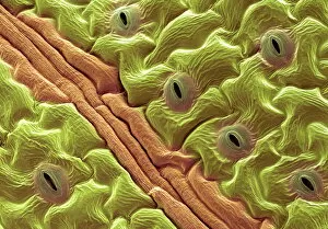
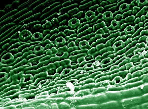
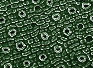
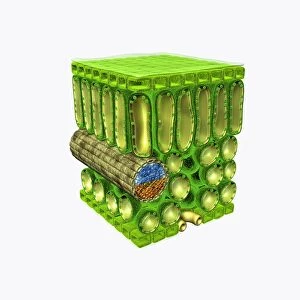
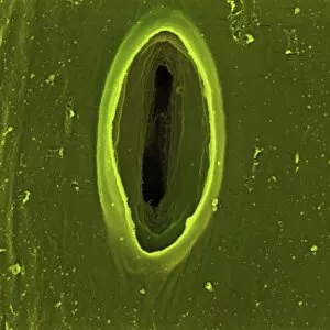
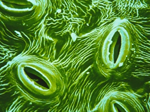
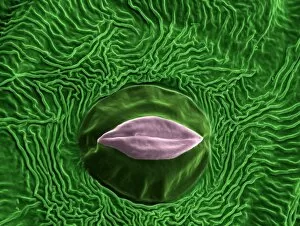
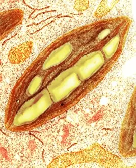
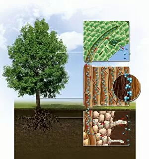
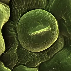
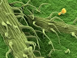
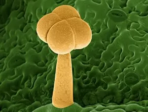
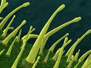
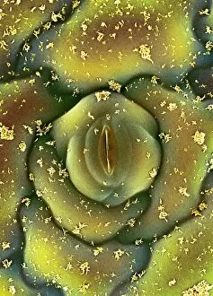
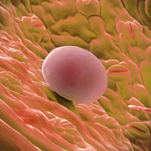
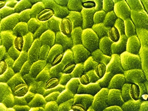
Stoma, also known as leaf pores, are fascinating structures found on the surface of plant leaves. These tiny openings play a crucial role in the exchange of gases between plants and their environment. In SEM (Scanning Electron Microscope) images such as Picture No. 11675585 and Picture No. 11675628, we can observe English oak leaf pores up close. The intricate patterns and shapes of these stomata highlight the complexity of nature's design. Moving on to other species, French lavender leaf pore captured in SEM reveals its unique characteristics that differ from those of English oak leaves. Similarly, Lemon grass and Chives leaf stoma (SEM C016 / 8060) showcase distinct features that make each plant species special. Coltsfoot leaf stomata (SEM C016 / 8052) provide us with another glimpse into the diverse world morphology. The variations in size, shape, and arrangement demonstrate how plants have adapted to different environmental conditions. Potato Leaf Stomata (SEM) exhibit yet another intriguing aspect structure. Their appearance suggests a specific adaptation strategy employed by potato plants for efficient gas exchange. Furthermore, studying stomata is not limited to individual cells but extends to understanding their distribution across an entire leaf surface like Elder leaves' epidermis with visible stomata under SEM observation. Lastly, illustrations depicting cross-sections through leaves offer valuable insights into the internal anatomy associated with stomatal function. Upper epidermis layers protect delicate palisade mesophyll tissues while facilitating gas diffusion through xylem vessels. Through these captivating images and illustrations, we gain a deeper appreciation for the intricate beauty and vital role played by stomata in sustaining life on our planet - enabling photosynthesis while regulating water loss through transpiration.