Fraud Blocker Skip to main content

Structural Collection (page 6)

"Unveiling the Intricate Web: Exploring Structural Wonders from Brain Fibres to DNA Molecules" Delving into the Complexity

Background imageStructural Collection: Picture No. 10897042

Picture No. 10897042
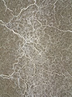
Structural forms of salt deposits at salt marshes in the central Death Valley Date:

Background imageStructural Collection: Picture No. 10897008

Picture No. 10897008
Structural forms at a dry pan in the Mesquite Flat Sand Dunes in the Death Valley Date:

Background imageStructural Collection: R H T Harper and A W Torry

R H T Harper and A W Torry
R.H.T. Harper, Chief Structural Engineer and A.W. Torry, Chief Stressman, de Havilland

Background imageStructural Collection: Martin XP6M-1 Seamaster during structural tests

Martin XP6M-1 Seamaster during structural tests in its Baltimore hangar

Background imageStructural Collection: Saunders-Roe A33 K4773

Saunders-Roe A33 K4773
Saunders-Roe A33, K4773, following structural failure of the mainplane during high-speed taxy tests in the Solent

Background imageStructural Collection: Multispectral scanner reveals lithologic and structural features of Great Namaland

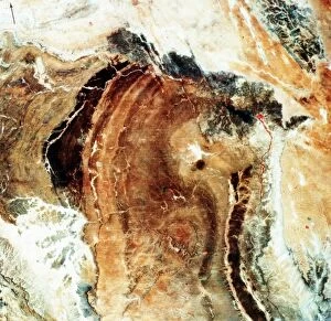
Multispectral scanner reveals lithologic and structural features of Great Namaland in Namibia with clarity. Area extremely dry with little vegetation, so geology dominates image

Background imageStructural Collection: Ricin A-chain, artwork C017 / 3653

Ricin A-chain, artwork C017 / 3653
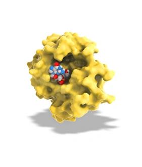
Ricin A-chain. Computer artwork showing the enzymatically active A-chain from a molecule of the toxic protein ricin. Ricin comprises two entwined amino acid chains; A (seen here) and B (not shown)

Background imageStructural Collection: Ricin molecule, artwork C017 / 3652

Ricin molecule, artwork C017 / 3652
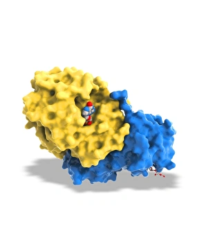
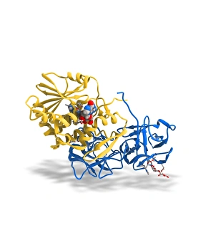
Ricin molecule. Computer artwork showing the structure of a molecule of the toxic protein ricin. Ricin comprises two entwined amino acid chains; A (yellow) and B (blue)
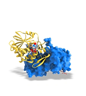
Background imageStructural Collection: Ricin molecule, artwork C017 / 3651

Ricin molecule, artwork C017 / 3651
Ricin molecule. Computer artwork showing the structure of a molecule of the toxic protein ricin. Ricin comprises two entwined amino acid chains; A (yellow) and B (blue)

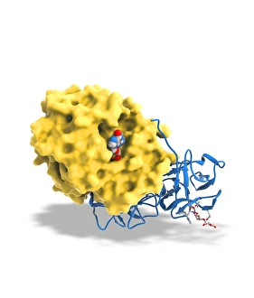
Background imageStructural Collection: Ricin molecule, artwork C017 / 3650

Ricin molecule, artwork C017 / 3650
Ricin molecule. Computer artwork showing the structure of a molecule of the toxic protein ricin. Ricin comprises two entwined amino acid chains; A (yellow) and B (blue)

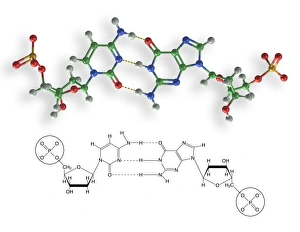
Background imageStructural Collection: Cytosine-guanine interaction, artwork C017 / 7215

Cytosine-guanine interaction, artwork C017 / 7215
Cytosine-guanine interaction. Computer artwork showing the structure of bound cytosine (left) and guanine molecules (right)

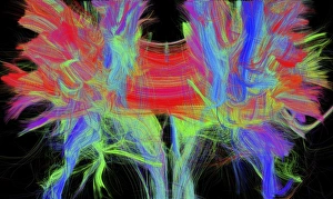
Background imageStructural Collection: Corpus callosum, DTI MRI scan C017 / 7044

Corpus callosum, DTI MRI scan C017 / 7044
Corpus callosum. Coronal 3D diffusion tensor imaging (DTI) magnetic resonance imaging (MRI) scan of nerve pathways (coloured) in and around the brains corpus callosum

Background imageStructural Collection: Cytosine-guanine interaction, artwork C017 / 7216

Cytosine-guanine interaction, artwork C017 / 7216
Cytosine-guanine interaction. Computer artwork showing the structure of bound cytosine (left) and guanine molecules (right)

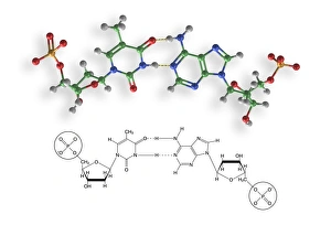
Background imageStructural Collection: Thymine-adenine interaction, artwork C017 / 7367

Thymine-adenine interaction, artwork C017 / 7367
Thymine-adenine interaction. Computer artwork showing the structure of bound thymine and adenine molecules. Atoms are shown as colour-coded spheres: carbon (green), hydrogen (white)

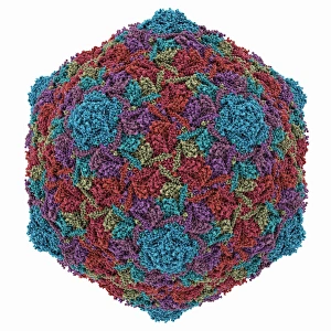
Background imageStructural Collection: HK97 bacteriophage capsid

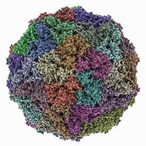
HK97 bacteriophage capsid, molecular model. Bacteriophages are viruses that infect bacteria, in this case enterobacteria such as E. coli (Escherichia coli), with the phage head shown here

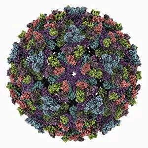
Background imageStructural Collection: Chikungunya virus capsid

Chikungunya virus capsid, molecular model. This virus, transmitted by mosquitoes in tropical Africa and Asia, causes fever and joint pain in humans, similar to dengue fever

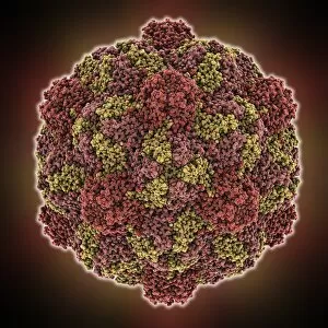
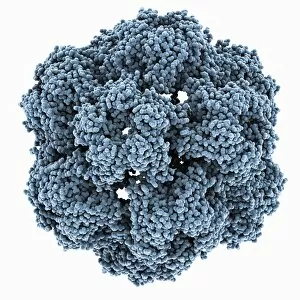
Background imageStructural Collection: HK97 bacteriophage procapsid

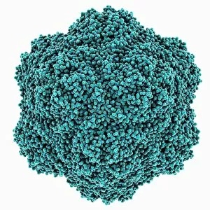
HK97 bacteriophage procapsid. Molecular model showing the structure of the prohead-I procapsid of the HK97 bacteriophage. Bacteriophages are viruses that infect bacteria

Background imageStructural Collection: Turnip yellow mosaic virus capsid

Turnip yellow mosaic virus capsid, molecular model. This virus infects a wide variety of plants, including crops such as turnips and cabbages, causing yellow patches on the leaves

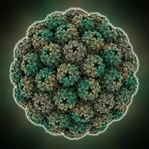
Background imageStructural Collection: Sindbis virus capsid, molecular model

Sindbis virus capsid, molecular model. This virus, transmitted by mosquitoes, causes sindbis fever in humans. In viruses, the capsid is the protein shell that encloses the genetic material

Background imageStructural Collection: Genetic research, conceptual image C014 / 1256

Genetic research, conceptual image C014 / 1256
Genetic research. Conceptual image of a molecular model of a strand of DNA (deoxyribonucleic acid) being held on a human hand

Background imageStructural Collection: Murine polyomavirus capsid

Murine polyomavirus capsid, molecular model. This virus, one of a range named for their potential to cause multiple tumours, infects mice

Background imageStructural Collection: Brome mosaic virus capsid

Brome mosaic virus capsid, molecular model. This plant virus infects grasses, especially brome grasses, and also barley. It causes mosaic patches of discolouration

Background imageStructural Collection: Cowpea chlorotic mottle virus capsid

Cowpea chlorotic mottle virus capsid, molecular model. This virus (CCMV) infects the cowpea plant (Vigna unguiculata), causing yellow spots of discolouration

Background imageStructural Collection: Potassium ion channel protein structure

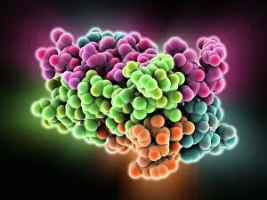
Potassium ion channel protein structure. Molecular model of a KcsA potassium ion (K+) channel from Streptomyces lividans bacteria

Background imageStructural Collection: Streptavidin-biotin molecular complex

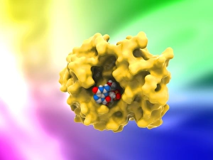
Streptavidin-biotin molecular complex. Molecular model of a single-strand binding complex of streptavidin (ribbons) and biotin (space-filled model, centre). Biotin is also known as vitamin B7

Background imageStructural Collection: Potassium ion channel beta subunit

Potassium ion channel beta subunit. Molecular model showing the structure a beta subunit of a voltage-dependent potassium (K+) channel

Background imageStructural Collection: KCNQ ion channel protein structure

KCNQ ion channel protein structure. Molecular model showing the protein structure of an ion channel domain. Ion channels are membrane-spanning proteins that form a pathway for the movement of

Background imageStructural Collection: Potassium ion channel cavity structure

Potassium ion channel cavity structure. Molecular model showing the structure of a cavity formed by potassium ion channel proteins

Background imageStructural Collection: Praseodymium, atomic structure

Praseodymium, atomic structure
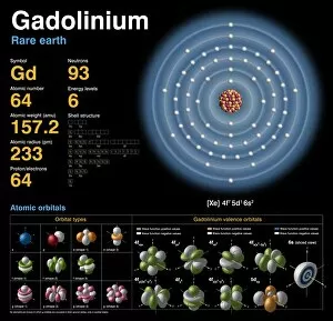
Bismuth (Bi). Diagram of the nuclear composition, electron configuration, chemical data, and valence orbitals of an atom of bismuth-209 (atomic number: 83), the most common isotope of this element

Background imageStructural Collection: Avian polyomavirus capsid

Avian polyomavirus capsid, molecular model. This virus, one of a range named for their potential to cause multiple tumours, infects birds. Discovered in budgerigars in 1981, it is often fatal

Background imageStructural Collection: Brain fibres, DTI MRI scan C017 / 7037

Brain fibres, DTI MRI scan C017 / 7037
Brain fibres. 3D diffusion tensor imaging (DTI) magnetic resonance imaging (MRI) scan of a selection of nerve pathways (green/yellow) in the brain

Background imageStructural Collection: Brain fibres, DTI MRI scan C017 / 7038

Brain fibres, DTI MRI scan C017 / 7038
Brain fibres. 3D diffusion tensor imaging (DTI) magnetic resonance imaging (MRI) scan of a selection of nerve pathways (green/yellow) in the brain

Background imageStructural Collection: Brain tumour, DTI MRI scan C017 / 7057

Brain tumour, DTI MRI scan C017 / 7057
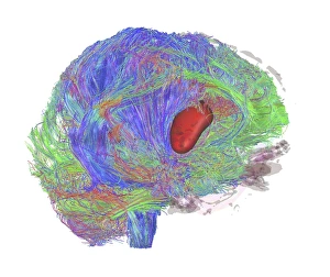
Brain tumour. 3D diffusion tensor imaging (DTI) magnetic resonance imaging (MRI) scan of nerve pathways (coloured) in a brain with a tumour (red, centre right)

Background imageStructural Collection: Cytoplasmic polyhedrosis virus capsid

Cytoplasmic polyhedrosis virus capsid, molecular model. Part of the Cypovirus genus and invariably fatal, this insect virus is transmitted by contamination of leaves eaten (examples include silkworms)

Background imageStructural Collection: Brain fibres, DTI MRI scan C017 / 7100

Brain fibres, DTI MRI scan C017 / 7100
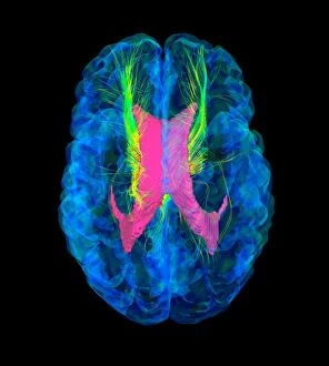
Brain fibres. 3D diffusion tensor imaging (DTI) magnetic resonance imaging (MRI) scan of a selection of nerve pathways (blue, pink, green) in the brain. The front of the brain is at left

Background imageStructural Collection: Theilers encephalomyelitis virus capsid

Theilers encephalomyelitis virus capsid, molecular model. This virus, which causes brain and spinal cord inflammation in mice, is used in research

Background imageStructural Collection: Brain tumour, DTI MRI scan C017 / 7059

Brain tumour, DTI MRI scan C017 / 7059
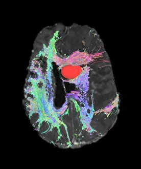
Brain tumour. Axial diffusion tensor imaging (DTI) magnetic resonance imaging (MRI) scan of nerve pathways (coloured) in a brain with a tumour (red, upper centre)

Background imageStructural Collection: Ricin A-chain, artwork C017 / 3654

Ricin A-chain, artwork C017 / 3654
Ricin A-chain. Computer artwork showing the enzymatically active A-chain from a molecule of the toxic protein ricin. Ricin comprises two entwined amino acid chains; A (seen here) and B (not shown)

Background imageStructural Collection: Brain tumour, DTI MRI scan C017 / 7058

Brain tumour, DTI MRI scan C017 / 7058
Brain tumour. 3D diffusion tensor imaging (DTI) magnetic resonance imaging (MRI) scan of nerve pathways (coloured) in a brain with a tumour (red, centre left)

Background imageStructural Collection: Phosphorus, atomic structure C018 / 3696

Phosphorus, atomic structure C018 / 3696
Argon (Ar). Diagram of the nuclear composition, electron configuration, chemical data, and valence orbitals of an atom of argon-40 (atomic number: 18)

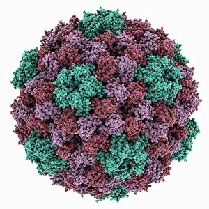
Background imageStructural Collection: Tobacco necrosis virus capsid

Tobacco necrosis virus capsid, molecular model. This plant virus infects a wide rage of plants, including the tobacco plant for which it is named. The virus causes tissue death (necrosis)

Background imageStructural Collection: Ricin molecule, artwork C017 / 3649

Ricin molecule, artwork C017 / 3649
Ricin molecule. Computer artwork showing the structure of a molecule of the toxic protein ricin. Ricin comprises two entwined amino acid chains; A (yellow) and B (blue)

Background imageStructural Collection: Grapevine fanleaf virus capsid

Grapevine fanleaf virus capsid, molecular model. This plant virus is named for its infection of grape vines. It is transmitted by the nematode worm Xiphinema index

Background imageStructural Collection: Cytosine molecule, artwork C017 / 7214

Cytosine molecule, artwork C017 / 7214
Cytosine molecule. Computer artwork showing the structure of a molecule of the nucleobase cytosine (2-oxy-4-aminopyrimidine)

Background imageStructural Collection: Transducin protein beta-gamma complex F006 / 9514

Transducin protein beta-gamma complex F006 / 9514
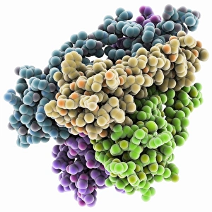
Transducin protein beta-gamma complex. Molecular model of the beta-gamma dimer of the heterotrimeric G protein transducin

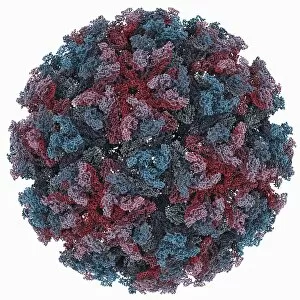
Background imageStructural Collection: Human poliovirus, molecular model F006 / 9434

Human poliovirus, molecular model F006 / 9434
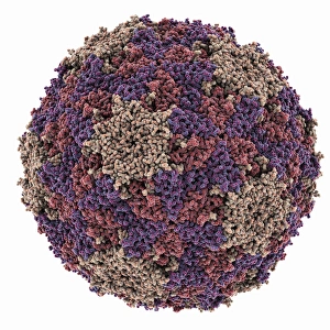
Human poliovirus particle. Molecular model of the capsid of the human poliovirus. The capsid is a protein coat that encloses the viruss genetic information (genome), stored as RNA (ribonucleic acid)

Background imageStructural Collection: VEE equine encephalitis virus capsid

VEE equine encephalitis virus capsid
Venezuelan equine encephalitis virus capsid, molecular model. This mosquito-borne virus can kill horses and other equine species, causing brain and spinal cord inflammation

Background imageStructural Collection: Sugar uptake in muscles, diagram

Sugar uptake in muscles, diagram
Sugar uptake in muscles. Diagram showing sugar from a drink (upper right) being taken up by the muscles of the human body


All Professionally Made to Order for Quick Shipping

"Unveiling the Intricate Web: Exploring Structural Wonders from Brain Fibres to DNA Molecules" Delving into the Complexity: DTI MRI scan C017 / 7099 reveals the intricate network of brain fibres, offering a glimpse into our cognitive architecture. Unraveling Mysteries: DTI MRI scan C017 / 7035 uncovers hidden connections within brain fibres, shedding light on how information flows through our neural pathways. Soaring Through History: The Hawker Hurricane Fighter, 1939 stands as a testament to the structural brilliance that revolutionized aerial warfare. Nature's Blueprint Revealed: A captivating light micrograph showcases the delicate structure of a dicotyledon plant stem, highlighting its role in supporting growth and nourishment. X-ray Artistry at its Finest: Skeletons come alive in mesmerizing artwork created through X-rays, capturing both their structural beauty and enigmatic allure. Building Blocks of Life: Dive deep into cellular wonders with an awe-inspiring glimpse into cell structures that form the foundation of all living organisms. Beneath the Surface Beauty: Witness skeletal marvels from below through stunning X-ray artwork that unveils hidden intricacies and celebrates human anatomy. Unlocking Genetic Secrets: A computer model presents a captivating visualization of DNA molecules, showcasing their structural elegance and vital role in heredity. Pollen's Dance in Light: Pine pollen grains take center stage under a microscope's gaze, revealing their exquisite architectural design crafted by nature itself. Illuminating Botanical Marvels: Explore the enchanting world within lime tree stems through a mesmerizing light micrograph that captures their unique structural patterns and resilience. Caffeine's Molecular Symphony : Journey inside caffeine's molecular realm as we uncover its intricate structure – fuel for millions around the globe.