Fraud Blocker Skip to main content

Structural Collection (page 9)

"Unveiling the Intricate Web: Exploring Structural Wonders from Brain Fibres to DNA Molecules" Delving into the Complexity

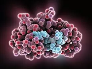
Background imageStructural Collection: Thymidylic acid-ribonuclease A complex

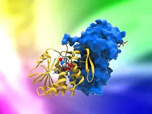
Thymidylic acid-ribonuclease A complex. Molecular model of a thymidylic acid tetramer (blue) in complex with ribonuclease A (red)

Background imageStructural Collection: DNA bundle on silicon nanopillars, SEM

DNA bundle on silicon nanopillars, SEM
DNA bundle on silicon nanopillars. Scanning electron micrograph (SEM) of a DNA (deoxyribonucleic acid) bundle and silicon nanopillars used to obtain the first high-contrast direct images of DNA

Background imageStructural Collection: MyoD muscle protein-DNA complex

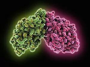
MyoD muscle protein-DNA complex. Molecular model of the MyoD muscle protein (green and blue) bound to a strand of DNA (deoxyribonucleic acid, orange and green)

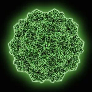
Background imageStructural Collection: Canine parvovirus capsid C015 / 8460

Canine parvovirus capsid C015 / 8460
Canine parvovirus capsid, molecular model. Canine parvovirus type 2 (CPV2) was first recognized in 1978 and spread worldwide in less than two years

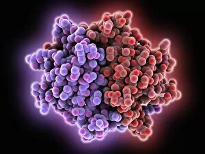
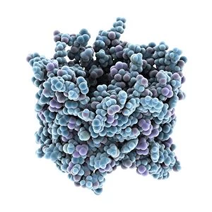
Background imageStructural Collection: VDAC-1 ion channel protein C015 / 8250

VDAC-1 ion channel protein C015 / 8250
VDAC-1 ion channel protein, molecular model. This is the human voltage-dependent anion-selective channel protein 1 (VDAC-1)

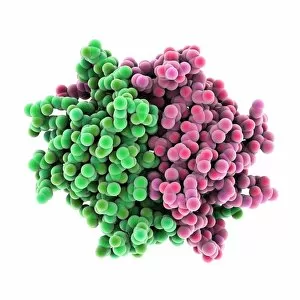
Background imageStructural Collection: Streptavidin bacterial protein C015 / 8458

Streptavidin bacterial protein C015 / 8458
Streptavidin bacterial protein, molecular model. Streptavidin is a protein obtained from the bacterium Streptomyces avidinii

Background imageStructural Collection: Calcium ATPase ion pump C015 / 7143

Calcium ATPase ion pump C015 / 7143
Calcium ATPase ion pump, molecular model. This enzyme is found in muscle cell membranes, where it pumps calcium in and out of muscle cells and controls muscle contractions

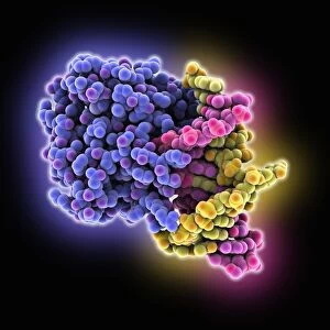
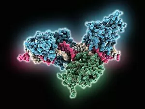
Background imageStructural Collection: DNA repair protein AlkB with DNA C016 / 0547

DNA repair protein AlkB with DNA C016 / 0547
DNA repair protein AlkB with DNA. Molecular model of the DNA (deoxyribonucleic acid) repair protein AlkB (blue) bound to a strand of double-stranded DNA (ds-DNA, pink and yellow)

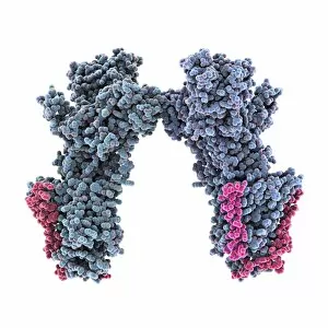
Background imageStructural Collection: Sodium-potassium ion pump proteins C015 / 9993

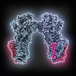
Sodium-potassium ion pump proteins C015 / 9993
Sodium-potassium ion pump proteins, molecular model. Sodium-potassium ATPase (adenosine triphosphatase) is an ATP-powered ion pump found in all animal cells

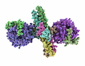
Background imageStructural Collection: Thrombin complexed with fibrinogen C015 / 7148

Thrombin complexed with fibrinogen C015 / 7148
Thrombin complexed with fibrinogen, molecular model. The thrombin molecules (left and right, purple and green) are bound to the central part of the fibrinogen molecule (centre, multiple colours)

Background imageStructural Collection: 3-hydroxyacyl-CoA dehydrogenase C015 / 9940

3-hydroxyacyl-CoA dehydrogenase C015 / 9940
3-hydroxyacyl-CoA dehydrogenase, molecular model. This enzyme is found in human heart tissue, and catalyzes a reaction that is part of the beta-oxidation pathway

Background imageStructural Collection: Streptavidin bacterial protein C015 / 8457

Streptavidin bacterial protein C015 / 8457
Streptavidin bacterial protein, molecular model. Streptavidin is a protein obtained from the bacterium Streptomyces avidinii

Background imageStructural Collection: Haemoglobin, molecular model C015 / 8938

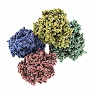
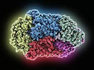
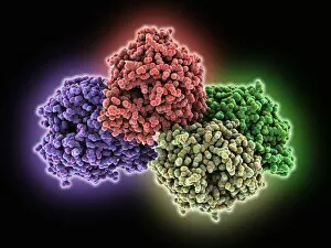
Haemoglobin, molecular model C015 / 8938
Haemoglobin, molecular model. This molecule transports oxygen around the body in red blood cells. It consists of four globin proteins (amino acid chains; orange, green, blue and purple)

Background imageStructural Collection: Sodium-potassium ion pump proteins C015 / 9997

Sodium-potassium ion pump proteins C015 / 9997
Sodium-potassium ion pump proteins, molecular model. Sodium-potassium ATPase (adenosine triphosphatase) is an ATP-powered ion pump found in all animal cells

Background imageStructural Collection: Retinal-producing oxygenase enzyme C015 / 7150

Retinal-producing oxygenase enzyme C015 / 7150
Retinal-producing oxygenase enzyme, molecular model. This enzyme, an oxygenase, plays a role in the production and metabolism of retinal and other apocarotenoids

Background imageStructural Collection: Central glycolytic gene regulator protein C016 / 0549

Central glycolytic gene regulator protein C016 / 0549
Central glycolytic gene regulator (CGGR) protein, molecular model. This protein binds to DNA (deoxyribonucleic acid) in the absence of glucose, blocking the transcription of certain genes

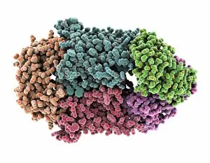
Background imageStructural Collection: Hemolysin-coregulated protein

Hemolysin-coregulated protein, molecular model. This protein is from the bacterium Pseudomonas aeruginosa. It is a hexameric ring structure, named hcp1

Background imageStructural Collection: Canine parvovirus capsid C015 / 8459

Canine parvovirus capsid C015 / 8459
Canine parvovirus capsid, molecular model. Canine parvovirus type 2 (CPV2) was first recognized in 1978 and spread worldwide in less than two years

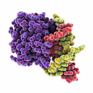
Background imageStructural Collection: DNA repair protein AlkB with DNA C016 / 0546

DNA repair protein AlkB with DNA C016 / 0546
DNA repair protein AlkB with DNA. Molecular model of the DNA (deoxyribonucleic acid) repair protein AlkB (purple) bound to a strand of double-stranded DNA (ds-DNA, red and green)

Background imageStructural Collection: Interferon-DNA transcription complex C015 / 8251

Interferon-DNA transcription complex C015 / 8251
Interferon-DNA transcription complex, molecular model. Bound to the DNA (deoxyribonucleic acid, green and yellow) is transcription factor p65, interferon regulatory factor 7

Background imageStructural Collection: Interferon-DNA transcription complex C015 / 8252

Interferon-DNA transcription complex C015 / 8252
Interferon-DNA transcription complex, molecular model. Bound to the DNA (deoxyribonucleic acid, pink and white) is transcription factor p65, interferon regulatory factor 7, interferon fusion protein

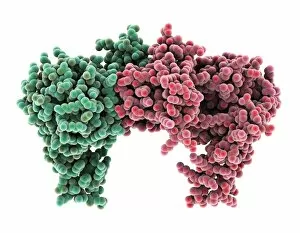
Background imageStructural Collection: Vitamin B12 import proteins C015 / 9942

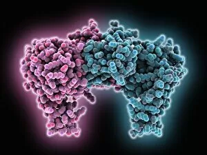
Vitamin B12 import proteins C015 / 9942
Vitamin B12 import proteins, molecular model. This complex is the import proteins btuC, btuD, and btuF. The first two together form BtuCD

Background imageStructural Collection: Haemoglobin, molecular model C015 / 8939

Haemoglobin, molecular model C015 / 8939
Haemoglobin, molecular model. This molecule transports oxygen around the body in red blood cells. It consists of four globin proteins (amino acid chains; orange, green, blue and purple)

Background imageStructural Collection: VDAC-1 ion channel protein C015 / 8249

VDAC-1 ion channel protein C015 / 8249
VDAC-1 ion channel protein, molecular model. This is the human voltage-dependent anion-selective channel protein 1 (VDAC-1)

Background imageStructural Collection: Thrombin complexed with fibrinogen C015 / 7149

Thrombin complexed with fibrinogen C015 / 7149
Thrombin complexed with fibrinogen, molecular model. The thrombin molecules (left and right, brown and pink) are bound to the central part of the fibrinogen molecule (centre, multiple colours)

Background imageStructural Collection: Simian virus (SV40) large T antigen C015 / 7069

Simian virus (SV40) large T antigen C015 / 7069
Simian virus (SV40) large T antigen, molecular model. This antigen is from the simian vacuolating virus 40 (SV40). Large T antigens play a role in regulating the viral life cycle of

Background imageStructural Collection: 3-hydroxyacyl-CoA dehydrogenase C015 / 9941

3-hydroxyacyl-CoA dehydrogenase C015 / 9941
3-hydroxyacyl-CoA dehydrogenase, molecular model. This enzyme is found in human heart tissue, and catalyzes a reaction that is part of the beta-oxidation pathway

Background imageStructural Collection: Vitamin B12 import proteins C015 / 9943

Vitamin B12 import proteins C015 / 9943
Vitamin B12 import proteins, molecular model. This complex is the import proteins btuC, btuD, and btuF. The first two together form BtuCD

Background imageStructural Collection: Calcium ATPase ion pump C015 / 7142

Calcium ATPase ion pump C015 / 7142
Calcium ATPase ion pump, molecular model. This enzyme is found in muscle cell membranes, where it pumps calcium in and out of muscle cells and controls muscle contractions

Background imageStructural Collection: Retinal-producing oxygenase enzyme C015 / 7151

Retinal-producing oxygenase enzyme C015 / 7151
Retinal-producing oxygenase enzyme, molecular model. This enzyme, an oxygenase, plays a role in the production and metabolism of retinal and other apocarotenoids

Background imageStructural Collection: Adeno-associated virus, molecular model C018 / 0449

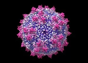
Adeno-associated virus, molecular model C018 / 0449
Adeno-associated virus (aV), molecular model. This image shows the core protein of the non-enveloped virus that surrounds the genetic material

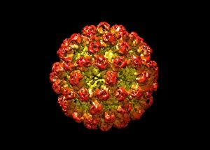
Background imageStructural Collection: Hepatitis B virus, molecular model C018 / 0455

Hepatitis B virus, molecular model C018 / 0455
Hepatitis B virus core protein, molecular model. The virus causes hepatitis B, an inflammatory liver disease. The core proteins enclose the virus DNA and are in turn surrounded by a lipid envelope
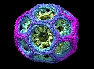
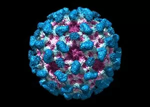
Background imageStructural Collection: Norovirus capsid, molecular model C018 / 0457

Norovirus capsid, molecular model C018 / 0457
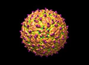
Norovirus capsid, molecular model. Also known as the winter vomiting bug, Noroviruses cause gastroenteritis and are highly contagious, infecting approximately 267 million people a year

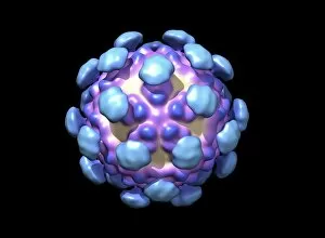
Background imageStructural Collection: Astrovirus capsid, molecular model C018 / 0450

Astrovirus capsid, molecular model C018 / 0450
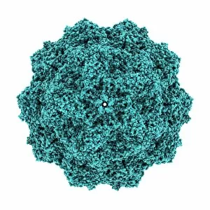
Astrovirus capsid, molecular model. This icosahedral virus was identified in 1975 using electron microscopy. It has a characteristic five-pointed symmetry to its surface, as seen here

Background imageStructural Collection: Hepatitis E virus, molecular model C018 / 0445

Hepatitis E virus, molecular model C018 / 0445
Hepatitis E virus core protein, molecular model. The virus causes hepatitis E, an inflammatory liver disease that usually only lasts a few weeks

Background imageStructural Collection: Human polio virus, molecular model

Human polio virus, molecular model
Human polio virus capsid, molecular model. Poliovirus causes poliomyelitis, a disease that can cause paralysis in up to 2 percent of patients, and in some cases death

Background imageStructural Collection: Clathrin lattice, molecular model C018 / 0453

Clathrin lattice, molecular model C018 / 0453
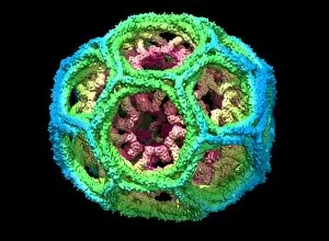
Clathrin lattice, molecular model. This polyhedral protein lattice coats eukaryotic cell membranes (vesicles) and is involved in protein secretion and membrane trafficking

Background imageStructural Collection: Clathrin lattice, molecular model C018 / 0452

Clathrin lattice, molecular model C018 / 0452
Clathrin lattice, molecular model. This polyhedral protein lattice coats eukaryotic cell membranes (vesicles) and is involved in protein secretion and membrane trafficking

Background imageStructural Collection: Clathrin lattice, molecular model C018 / 0454

Clathrin lattice, molecular model C018 / 0454
Clathrin lattice, molecular model. This polyhedral protein lattice coats eukaryotic cell membranes (vesicles) and is involved in protein secretion and membrane trafficking

Background imageStructural Collection: KSHV virus capsid, molecular model C018 / 0456

KSHV virus capsid, molecular model C018 / 0456
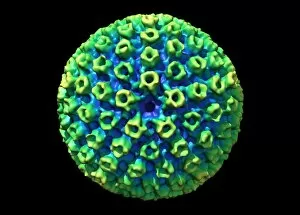
KSHV virus capsid, molecular model. KSHV is Kaposis sarcoma-associated herpesvirus. The virus is an oncovirus, which is a virus that can cause cancer

Background imageStructural Collection: Corpus callosum, DTI MRI scan C017 / 7045

Corpus callosum, DTI MRI scan C017 / 7045
Corpus callosum. Coronal 3D diffusion tensor imaging (DTI) magnetic resonance imaging (MRI) scan of nerve pathways (blue) in and around the brains corpus callosum

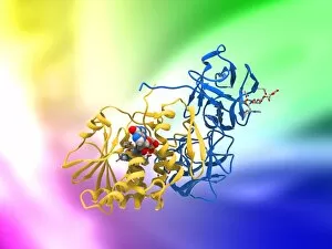
Background imageStructural Collection: Ricin molecule, artwork C017 / 3656

Ricin molecule, artwork C017 / 3656
Ricin molecule. Computer artwork showing the structure of a molecule of the toxic protein ricin. Ricin comprises two entwined amino acid chains; A (yellow) and B (blue)

Background imageStructural Collection: Ricin molecule, artwork C017 / 3655

Ricin molecule, artwork C017 / 3655
Ricin molecule. Computer artwork showing the structure of a molecule of the toxic protein ricin. Ricin comprises two entwined amino acid chains; A (yellow) and B (blue)

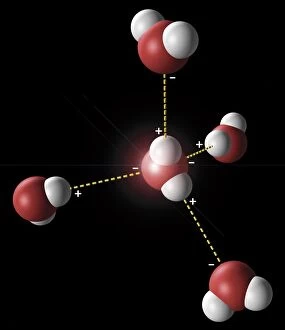
Background imageStructural Collection: Hydrogen bonding in water, artwork C018 / 3560

Hydrogen bonding in water, artwork C018 / 3560
Hydrogen bonding in water. Artwork showing the hydrogen bonding (yellow dotted lines) between five water molecules. There is a weak, partial negative charge on the oxygen atoms (red)

Background imageStructural Collection: Guanine molecule, artwork C017 / 7239

Guanine molecule, artwork C017 / 7239
Guanine molecule. Computer artwork showing the structure of a molecule of the nucleobase guanine. Atoms are shown as colour-coded spheres: carbon (green), hydrogen (white)

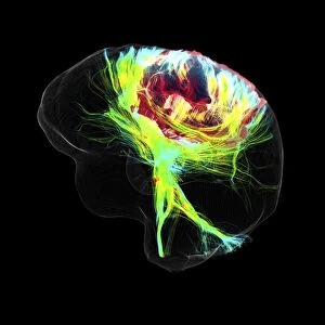
Background imageStructural Collection: Glioblastoma brain tumour, DTI MRI scan C017 / 7052

Glioblastoma brain tumour, DTI MRI scan C017 / 7052
Glioblastoma brain tumour. 3D diffusion tensor imaging (DTI) magnetic resonance imaging (MRI) scan of nerve pathways (coloured) around a glioblastoma (upper right) in the brain

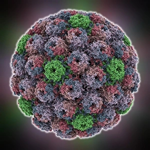
Background imageStructural Collection: SV40 virus capsid, molecular model

SV40 virus capsid, molecular model. Simian virus 40 (SV40) is found in monkeys such as Rhesus monkeys and macaques. Potentially tumour-causing, it is used in laboratory research and in vaccines

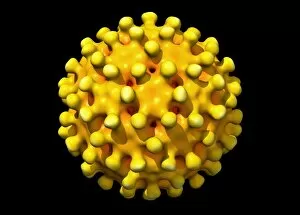
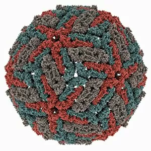
Background imageStructural Collection: Dengue virus capsid, molecular model

Dengue virus capsid, molecular model. This virus, transmitted by mosquito bites, causes the tropical disease dengue fever in humans


All Professionally Made to Order for Quick Shipping

"Unveiling the Intricate Web: Exploring Structural Wonders from Brain Fibres to DNA Molecules" Delving into the Complexity: DTI MRI scan C017 / 7099 reveals the intricate network of brain fibres, offering a glimpse into our cognitive architecture. Unraveling Mysteries: DTI MRI scan C017 / 7035 uncovers hidden connections within brain fibres, shedding light on how information flows through our neural pathways. Soaring Through History: The Hawker Hurricane Fighter, 1939 stands as a testament to the structural brilliance that revolutionized aerial warfare. Nature's Blueprint Revealed: A captivating light micrograph showcases the delicate structure of a dicotyledon plant stem, highlighting its role in supporting growth and nourishment. X-ray Artistry at its Finest: Skeletons come alive in mesmerizing artwork created through X-rays, capturing both their structural beauty and enigmatic allure. Building Blocks of Life: Dive deep into cellular wonders with an awe-inspiring glimpse into cell structures that form the foundation of all living organisms. Beneath the Surface Beauty: Witness skeletal marvels from below through stunning X-ray artwork that unveils hidden intricacies and celebrates human anatomy. Unlocking Genetic Secrets: A computer model presents a captivating visualization of DNA molecules, showcasing their structural elegance and vital role in heredity. Pollen's Dance in Light: Pine pollen grains take center stage under a microscope's gaze, revealing their exquisite architectural design crafted by nature itself. Illuminating Botanical Marvels: Explore the enchanting world within lime tree stems through a mesmerizing light micrograph that captures their unique structural patterns and resilience. Caffeine's Molecular Symphony : Journey inside caffeine's molecular realm as we uncover its intricate structure – fuel for millions around the globe.