
T Cell Collection
"T cells: The Mighty Warriors Against Cancer and HIV" T lymphocytes, also known as T cells, are a type of white blood cell that play a crucial role in our immune system


All Professionally Made to Order for Quick Shipping
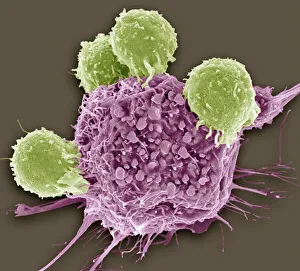
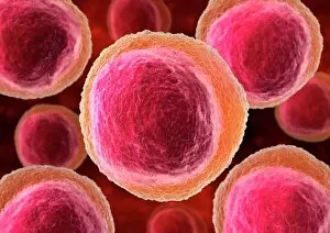
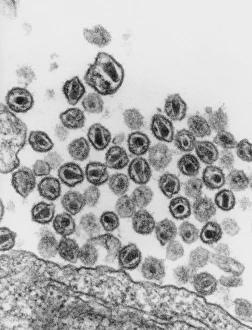
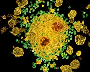
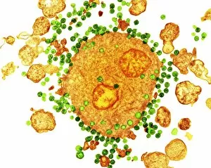
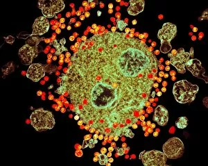
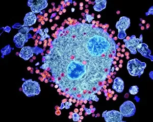
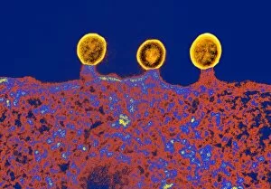
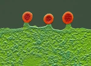
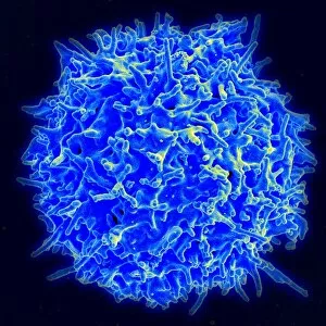
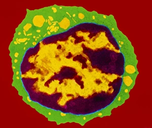
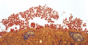
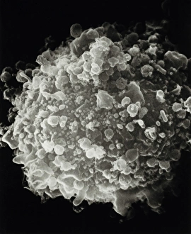
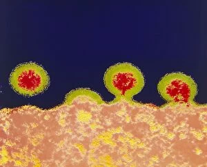
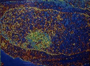
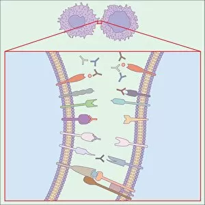
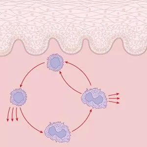
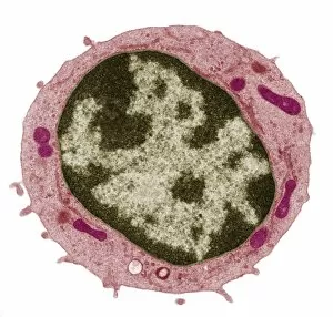
"T cells: The Mighty Warriors Against Cancer and HIV" T lymphocytes, also known as T cells, are a type of white blood cell that play a crucial role in our immune system. These remarkable warriors have the power to recognize and destroy cancer cells, as well as combat the notorious human immunodeficiency virus (HIV). In an astonishing SEM C001 / 1679 image, we witness the magnificence of T lymphocytes engaging with cancer cells. Their intricate interactions hold immense potential for targeted therapies against this devastating disease. Artwork depicting lymphocyte white blood cells showcases their elegance and importance in safeguarding our health. These tiny soldiers tirelessly patrol our bodies, ready to defend us from any foreign invaders. TEM images reveal the harsh reality of HIV viruses budding from T-cells. In these haunting visuals (such as TEM C018 / 8599), we witness the battle between our immune system's guardians and this relentless virus that causes AIDS. Yet even amidst adversity, hope remains. Astonishingly captured by TEM techniques (C018 / 0125-0127), budding HIV particles showcase both the resilience of these viruses and the determination of T-cells fighting back against them, and is through understanding these complex dynamics that scientists strive towards finding effective treatments or even a cure for this global epidemic. Furthermore, another captivating image shows a T-cell receptor bound to enterotoxin—a reminder of how intricately designed our immune defense mechanisms truly are. This interaction highlights not only their ability to identify harmful substances but also their potential for therapeutic interventions. As research continues to unravel more about T-cells' incredible capabilities, we gain insights into new avenues for combating diseases like cancer and HIV/AIDS. With each breakthrough discovery represented by these stunning images, we move closer towards harnessing the full potential of these mighty warriors within us all – our very own T cells.