
T Lymphocyte Collection
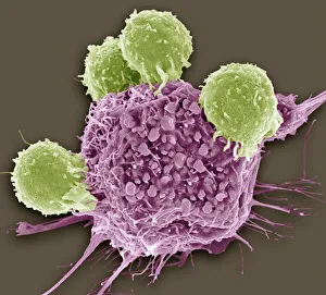
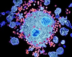
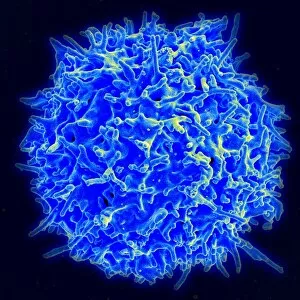
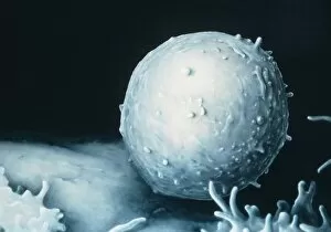
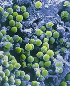
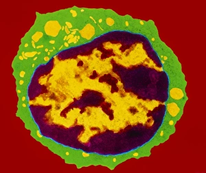
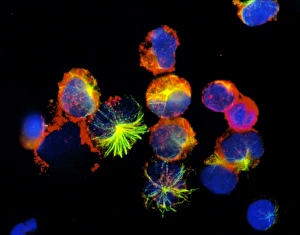
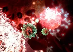
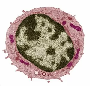
T lymphocytes, also known as T cells, play a crucial role in the immune system's defense against cancer cells


All Professionally Made to Order for Quick Shipping
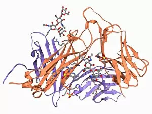
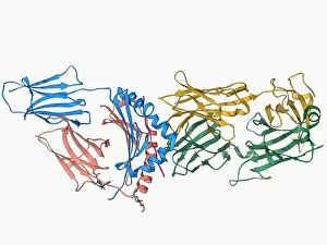
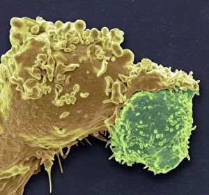
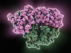
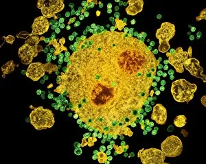
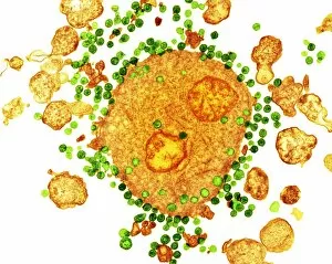
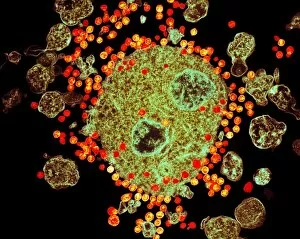
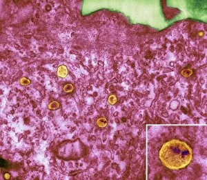
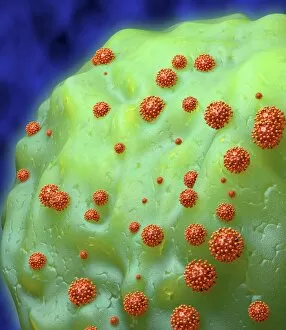
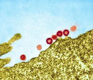
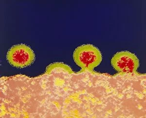
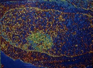
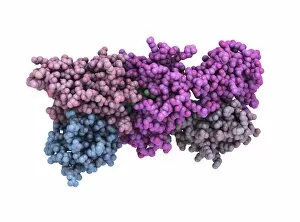
T lymphocytes, also known as T cells, play a crucial role in the immune system's defense against cancer cells. In SEM C001 / 1679 image, we can observe these remarkable blood cells in action. Another fascinating view captured by TEM reveals AIDS viruses budding from a cell, highlighting the importance of T lymphocytes in combating viral infections like HIV. Molecular models such as the T cell receptor (F006 / 9515) and its complex with antigens (F006 / 9339) shed light on how T lymphocytes recognize foreign invaders. The intricate structure of the T-cell receptor B7 molecule (F006 / 9247) further emphasizes their specificity and efficiency in targeting threats to our health. One captivating image shows the binding between a T-cell receptor and an enterotoxin, illustrating how these receptors are capable of recognizing harmful substances and initiating immune responses. This interaction is vital for protecting our bodies from toxins that could cause harm. Moreover, white blood cell antigen presentation images (C016 / 9058 & C016 / 9057) showcase another critical function of T lymphocytes – presenting antigens to other immune cells for coordinated attacks against pathogens or abnormal cells.