Fraud Blocker Skip to main content

Thigh Bone Collection

"The Thigh Bone: A Journey of Art, Replacement, and Resilience" Hip replacement meets artistry - a fusion of medical innovation and creative expression

Background imageThigh Bone Collection: Hip replacement, artwork

Hip replacement, artwork
Hip replacement. Artwork of a total hip replacement. The prosthetic hip joint consists of a metal shaft and ball (pink) attached to the top of the femur (thigh bone) and a plastic socket (white)

Background imageThigh Bone Collection: Total hip replacement, X-ray

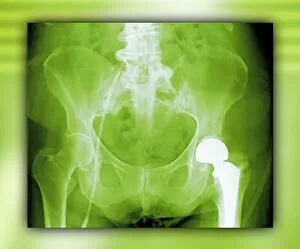
Total hip replacement, X-ray
Total hip replacement. Coloured frontal X-ray of a female pelvis with a total hip replacement (white, lower right). The replacement hip consists of a ball and shaft

Background imageThigh Bone Collection: Damaged knee ligament, artwork

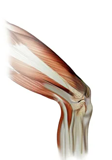
Damaged knee ligament, artwork
Damaged knee ligament. Computer artwork of the muscle structure of the leg and anatomy of the human knee joint, showing a tear (dark line, centre right) in one of the cruciate ligaments (beige)

Background imageThigh Bone Collection: Healthy knee, X-ray

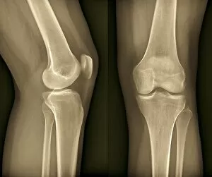
Healthy knee, X-ray
Healthy knee. Coloured frontal (right) and profile (left) X-rays of the healthy knee of a 24 year old patient

Background imageThigh Bone Collection: Human Skeleton (engraving)

Human Skeleton (engraving)
2802021 Human Skeleton (engraving) by English School, (19th century); Private Collection; (add.info.: Human Skeleton. Illustration for An Illustrated Vocabulary For The Use Of The Deaf And Dumb)

Background imageThigh Bone Collection: Normal legs, X-rays

Normal legs, X-rays
Normal legs. Coloured frontal X-rays of the healthy legs, of a 35-year-old

Background imageThigh Bone Collection: Healthy knee, X-ray F006 / 9125

Healthy knee, X-ray F006 / 9125
Healthy knee. Coloured frontal (right) and profile (left) X-rays of the healthy knee of a 24 year old patient

Background imageThigh Bone Collection: Healthy knee, CT scan C018 / 0413

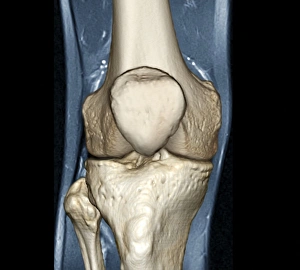
Healthy knee, CT scan C018 / 0413
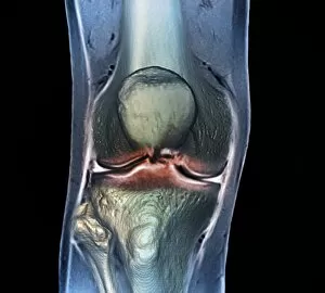
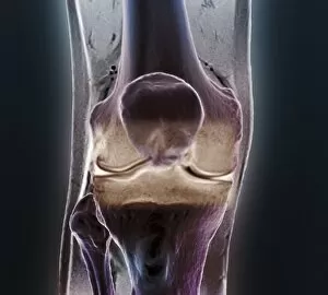
Healthy knee. Coloured frontal computed tomography (CT) scan projected on to a magnetic resonance imaging (MRI) scan of the knee of a 30 year old

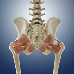
Background imageThigh Bone Collection: Buttock muscles, artwork C013 / 4414

Buttock muscles, artwork C013 / 4414
Buttock muscles. Computer artwork of a posterior view of the hips showing some of the muscles (red) of the buttocks. These are shown attached to the femurs (thigh bones) and the pelvis bones

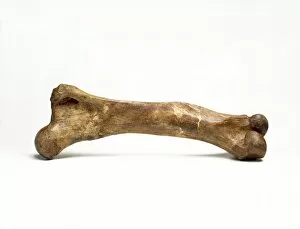
Background imageThigh Bone Collection: Mammoth thigh bone

Mammoth thigh bone
Fossilised thigh bone of a woolly mammoth (Mammuthus primigenius) from Siberia. About 40, 000 years old

Background imageThigh Bone Collection: Illustration of human bones, femur

Illustration of human bones, femur
Medicine: Human anatomy, bones, femur

Background imageThigh Bone Collection: Using kitchen knife to remove bone from chicken thigh

Using kitchen knife to remove bone from chicken thigh

Background imageThigh Bone Collection: Osteoarthritis of the hip, X-ray F006 / 3744

Osteoarthritis of the hip, X-ray F006 / 3744
Osteoarthritis of the hip. Coloured frontal X-ray (front view) of the pelvis in a 59 year old patient showing osteoarthritis of both hip joints

Background imageThigh Bone Collection: Osteoarthritis of the hip, X-ray F006 / 3745

Osteoarthritis of the hip, X-ray F006 / 3745
Osteoarthritis of the hip. Coloured frontal X-ray (front view) of the pelvis in a 59 year old patient showing osteoarthritis of both hip joints

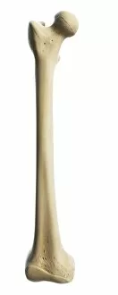
Background imageThigh Bone Collection: Thigh bone, artwork F007 / 7063

Thigh bone, artwork F007 / 7063
Thigh (femur) bone, computer artwork

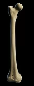
Background imageThigh Bone Collection: Thigh bone, artwork F007 / 7058

Thigh bone, artwork F007 / 7058
Thigh (femur) bone, computer artwork

Background imageThigh Bone Collection: Thigh bone, artwork F007 / 7057

Thigh bone, artwork F007 / 7057
Thigh (femur) bone, computer artwork

Background imageThigh Bone Collection: Thigh bone, artwork F007 / 7060

Thigh bone, artwork F007 / 7060
Thigh (femur) bone, computer artwork

Background imageThigh Bone Collection: Thigh bone, artwork F007 / 7061

Thigh bone, artwork F007 / 7061
Thigh (femur) bone, computer artwork

Background imageThigh Bone Collection: Thigh bone, artwork F007 / 7062

Thigh bone, artwork F007 / 7062
Thigh (femur) bone, computer artwork

Background imageThigh Bone Collection: Thigh bone, artwork F007 / 7059

Thigh bone, artwork F007 / 7059
Thigh (femur) bone, computer artwork

Background imageThigh Bone Collection: Thigh bone, artwork F006 / 3070

Thigh bone, artwork F006 / 3070
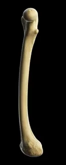
Thigh bone. Computer artwork showing the femur bone

Background imageThigh Bone Collection: Thigh bone, artwork F006 / 2997

Thigh bone, artwork F006 / 2997
Thigh bone. Computer artwork showing the femur bone

Background imageThigh Bone Collection: Deep abdominal organs, 1880 artwork C017 / 6913

Deep abdominal organs, 1880 artwork C017 / 6913
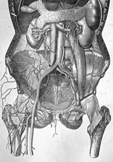
Deep abdominal organs. Historical anatomical artwork of a dissected abdomen seen from the front, showing the deep-seated organs and major blood vessels after the removal of the intestines

Background imageThigh Bone Collection: Hip pain, artwork C013 / 5917

Hip pain, artwork C013 / 5917
Hip pain, computer artwork

Background imageThigh Bone Collection: Malunited fracture, X-ray C017 / 7640

Malunited fracture, X-ray C017 / 7640
Malunited fracture. X-ray of the leg of a 64 year old female patient with a malunited fracture of the femur (thigh bone). This occurs where a fracture fails to heal in the correct position

Background imageThigh Bone Collection: Arthritis of the knee, X-ray F006 / 9104

Arthritis of the knee, X-ray F006 / 9104
Arthritis of the knee. Coloured X-ray of the knee of an obese 48 year old patient showing degenerative arthritis caused by the extreme stress on the knee from being overweight

Background imageThigh Bone Collection: Arthritis of the knee, X-ray F006 / 9103

Arthritis of the knee, X-ray F006 / 9103
Arthritis of the knee. Coloured X-ray of the knee of an obese 48 year old patient showing degenerative arthritis caused by the extreme stress on the knee from being overweight

Background imageThigh Bone Collection: Normal knees, X-rays

Normal knees, X-rays
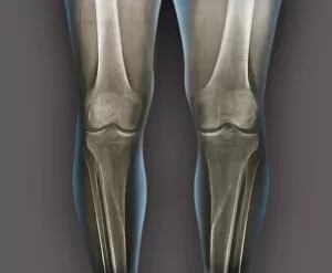
Normal knees. Coloured frontal X-rays of the healthy knees of a 35-year-old

Background imageThigh Bone Collection: Total knee replacement, X-ray

Total knee replacement, X-ray
Total knee replacement. X-ray of the legs of a 49-year-old patient who has had a total knee replacement (white) of the left knee (right)

Background imageThigh Bone Collection: Hip bones, artwork C016 / 7015

Hip bones, artwork C016 / 7015
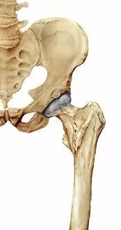
Hip bones. Artwork of a frontal (anterior) view of the left hip, showing the bones that articulate to form this joint. At lower right is the femur (thigh bone)
Background imageThigh Bone Collection: Arthritis of the knee, 3D CT scan C016 / 6510

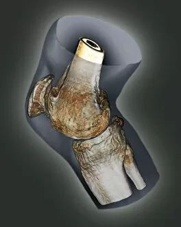
Arthritis of the knee, 3D CT scan C016 / 6510
Arthritis of the knee. Coloured 3D computed tomography (CT) scan of the right knee of a 65 year old patient with severe osteoarthritis

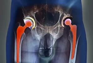
Background imageThigh Bone Collection: Double hip replacement, X-ray C016 / 6447

Double hip replacement, X-ray C016 / 6447
Double hip replacement. Coloured X-ray of the pelvis of a 63 year old male patient who has had two total hip replacements

Background imageThigh Bone Collection: Knee bones and ligaments, artwork C016 / 7012

Knee bones and ligaments, artwork C016 / 7012
Knee bones and ligaments. Artwork of a frontal view of the knee joint, showing bones, ligaments, tendons, and cartilage. The bones are the femur (thigh bone, top), patella (knee-cap, upper centre)

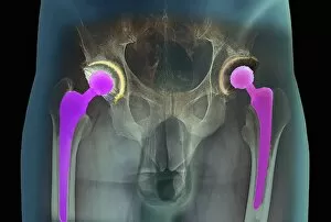
Background imageThigh Bone Collection: Double hip replacement, X-ray C016 / 6448

Double hip replacement, X-ray C016 / 6448
Double hip replacement. Coloured X-ray of the pelvis of a 63 year old male patient who has had two total hip replacements

Background imageThigh Bone Collection: Arthritis of the knee, 3D CT scan C016 / 6511

Arthritis of the knee, 3D CT scan C016 / 6511
Arthritis of the knee. Coloured 3D computed tomography (CT) scan of the right knee of a 65 year old patient with severe osteoarthritis

Background imageThigh Bone Collection: Double hip replacement, X-ray C016 / 6446

Double hip replacement, X-ray C016 / 6446
Double hip replacement. Coloured X-ray of the pelvis of a 63 year old male patient who has had two total hip replacements

Background imageThigh Bone Collection: Knee bones and ligaments, artwork C016 / 7011

Knee bones and ligaments, artwork C016 / 7011
Knee bones and ligaments. Artwork of a frontal view of the knee joint, showing bones, ligaments, tendons and cartilage. The bones are the femur (thigh bone, top), patella (knee-cap, upper centre)

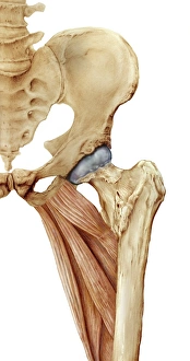
Background imageThigh Bone Collection: Hip bones and thigh muscles, artwork C016 / 7016

Hip bones and thigh muscles, artwork C016 / 7016
Hip bones and thigh muscles, artwork

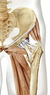
Background imageThigh Bone Collection: Hip bones, muscles and ligaments, artwork C016 / 7018

Hip bones, muscles and ligaments, artwork C016 / 7018
Hip bones and thigh muscles. Artwork of a frontal (anterior) view of the left hip, showing bones and some thigh muscles (red). At lower right is the femur (thigh bone)

Background imageThigh Bone Collection: Healthy knee, CT scan C018 / 0415

Healthy knee, CT scan C018 / 0415
Healthy knee. Coloured frontal computed tomography (CT) scan projected on to a magnetic resonance imaging (MRI) scan of the knee of a 30 year old

Background imageThigh Bone Collection: Healthy knee, CT scan C018 / 0414

Healthy knee, CT scan C018 / 0414
Healthy knee. Coloured frontal computed tomography (CT) scan projected on to a magnetic resonance imaging (MRI) scan of the knee of a 30 year old

Background imageThigh Bone Collection: Knee injury, 3D CT scan C018 / 0648

Knee injury, 3D CT scan C018 / 0648
Knee injury. Coloured 3D computed tomography (CT) scan of a knee with a meniscal tear (white line at right between bones)

Background imageThigh Bone Collection: 17 month old babys pelvis, X-ray

17 month old babys pelvis, X-ray
Seventeen month old babys pelvis, coloured X-ray. The hip joints (centre right and centre left) are still forming

Background imageThigh Bone Collection: Osteoarthritis of the hip, X-ray C018 / 0593

Osteoarthritis of the hip, X-ray C018 / 0593
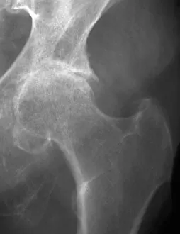
Osteoarthritis of the hip. X-ray the left hip of a 74 year old patient showing osteoarthritis. The ball at the top of the femur (thigh bone) fits into the socket of the hip

Background imageThigh Bone Collection: Knee injury, 3D CT scan C018 / 0639

Knee injury, 3D CT scan C018 / 0639
Knee injury. Coloured 3D computed tomography (CT) scan of a knee with a meniscal tear (indicated by arrows). The menisci are fibrocartilage crescents that act as shock absorbers between the femur

Background imageThigh Bone Collection: Rickets, illustration C018 / 0779

Rickets, illustration C018 / 0779
Rickets. Illustration of a femur (thigh bone) that has become curved due to rickets. Rickets, a softening of the bones, is caused by a lack of vitamin D

Background imageThigh Bone Collection: Knee injury, 3D CT scan C018 / 0645

Knee injury, 3D CT scan C018 / 0645
Knee injury. Coloured 3D computed tomography (CT) scan of a knee with a meniscal tear (blue line at right between bones). The menisci are fibrocartilage crescents that act as shock absorbers between


All Professionally Made to Order for Quick Shipping

"The Thigh Bone: A Journey of Art, Replacement, and Resilience" Hip replacement meets artistry - a fusion of medical innovation and creative expression. Peering into the depths of healing - an X-ray reveals the success of total hip replacement surgery. From injury to inspiration - damaged knee ligament transformed into captivating artwork. Unveiling strength within - a healthy knee captured in its intricate beauty through an X-ray lens. Exploring the human frame - an enchanting engraving depicting the timeless elegance of our skeleton. Beneath the surface lies perfection - normal legs revealed through mesmerizing X-rays. Literary musings on anatomy's marvels - Henry Fielding's illustration brings Tom Jones to life alongside a femur bone engraving. Delving deeper for answers – CT scan captures the essence of a healthy knee in stunning detail (CT scan C018 / 0413). Celebrating power from behind – artistic portrayal showcases buttock muscles' strength and grace (artwork C013 / 4414). The foundation of our being unveiled – an exquisite illustration showcasing the majesty of human bones, including the mighty femur. Culinary exploration meets anatomy – using a kitchen knife to remove bone from chicken thigh highlights similarities between nature and ourselves. Osteoarthritis unraveled–an X-ray revealing F006/3744 showcases how it affects one's hip joint.