
Unicellular Collection
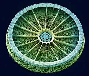
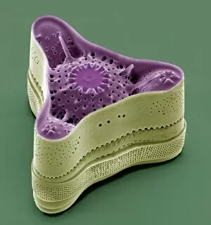
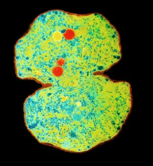
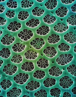
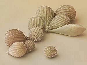
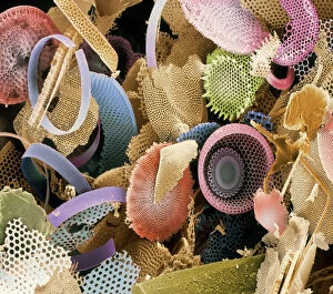
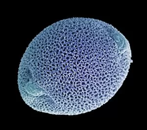
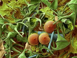
Unicellular organisms, such as diatoms and blue-green algae, are fascinating to explore under the scanning electron microscope (SEM


All Professionally Made to Order for Quick Shipping
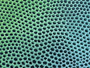
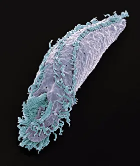
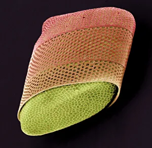
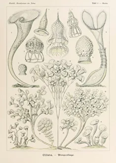
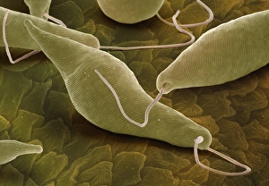
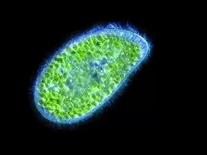
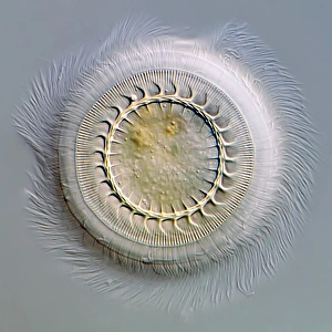
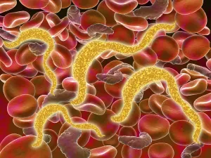
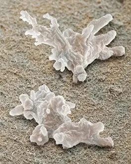
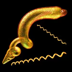
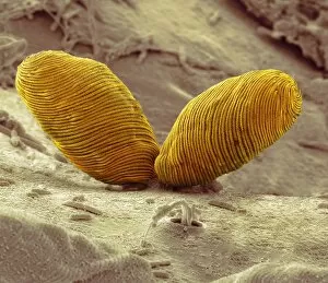
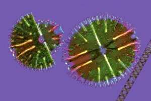
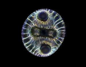
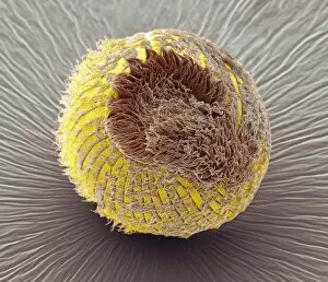
Unicellular organisms, such as diatoms and blue-green algae, are fascinating to explore under the scanning electron microscope (SEM). Their intricate structures and diverse forms never cease to amaze scientists. Take a closer look at these microscopic wonders. In the world of diatoms, their beauty lies in their silica-based cell walls. Under SEM, we can observe the delicate patterns etched onto their frustules, showcasing nature's artistic touch. Microcystis blue-green alga is another unicellular organism that catches our attention. Its vibrant blue color stands out amidst other microscopic lifeforms. SEM allows us to appreciate its unique shape and structure up close. Moving on to Acrosphaera radiolarian, a type organism known for its intricate skeletal structure resembling a miniature work of art. SEM reveals every tiny detail of this beautiful creature. Even in ancient times, diatoms left behind fossilized remains that continue to captivate researchers today. Through SEM imaging, we can witness these preserved diatom shells from ages past and gain insights into Earth's history. Dinoflagellates are yet another group organisms worth exploring under SEM. These remarkable creatures possess flagella that enable them to move with grace through water bodies while displaying stunning shapes and patterns when magnified. Let's not forget about Oxytricha ciliate protozoan. This peculiar-looking creature exhibits an array of hair-like projections called cilia which aid in locomotion and feeding. Thanks to SEM technology, we can marvel at its intricately structured body. Unicellular lifeforms like diatom algae have so much more than meets the eye – or even what traditional microscopes reveal. With SEM imaging techniques available today, scientists uncover hidden details within these tiny organisms that contribute immensely to our understanding of biodiversity and evolution. So next time you come across a mention of "unicellular, " remember the vast world waiting beneath the surface, waiting to be explored through SEM.