
Ureter Collection
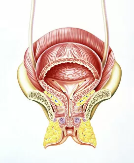
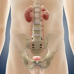
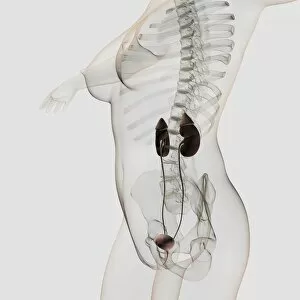
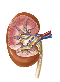
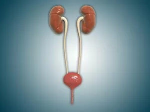
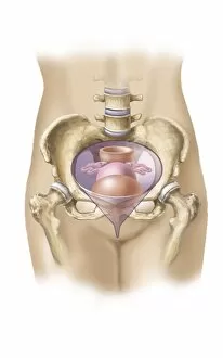
The ureter, a vital component of the female urinary bladder, is beautifully depicted in this artwork showcasing the intricate internal anatomy


All Professionally Made to Order for Quick Shipping
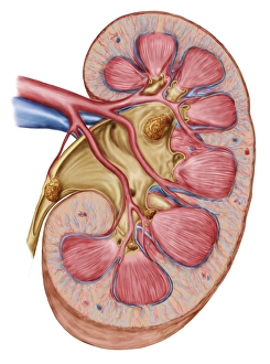
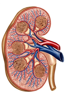
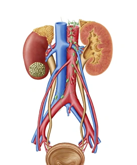
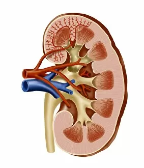
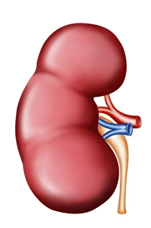
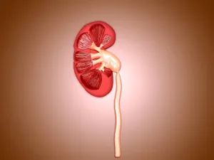
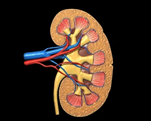
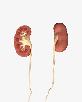
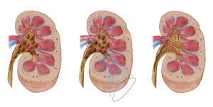
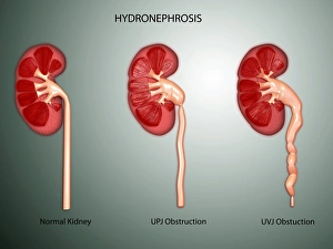
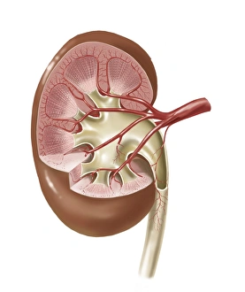
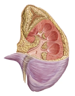
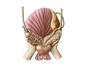
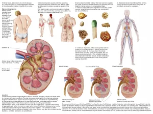
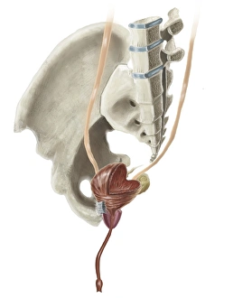
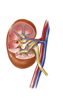
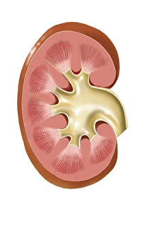
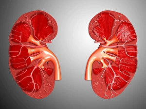
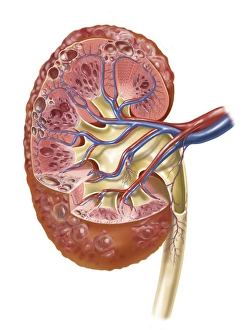
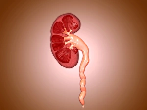
The ureter, a vital component of the female urinary bladder, is beautifully depicted in this artwork showcasing the intricate internal anatomy. The detailed section through the female bladder reveals its connection to the kidneys, which are also showcased in stunning artwork. The kidney's complex structure and function are brought to life through these illustrations, providing a deeper understanding of their importance in our bodies. Not limited to human anatomy alone, this collection includes artwork depicting dog and deer anatomy as well. A cross-section of the internal anatomy of the kidney allows us to explore its inner workings further. However, it is crucial to note that not all conditions related to this organ are benign. An early stage of kidney cancer is visually represented with a visible tumor on one side of the kidney. This serves as a reminder that vigilance and regular check-ups are essential for maintaining good health. Lastly, we have an artwork dedicated to male urinary system highlighting how both genders share similar structures but differ in certain aspects.