
Vector Borne Collection
"Unveiling the World of Vector-Borne Diseases: A Microscopic Journey" Step into the microscopic realm and explore the intricate world of vector-borne diseases


All Professionally Made to Order for Quick Shipping
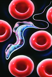
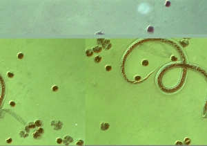
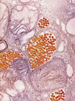
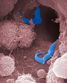
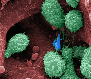
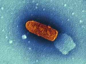
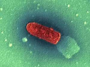
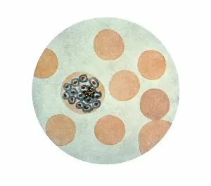
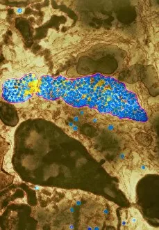
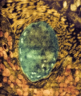
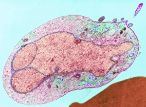
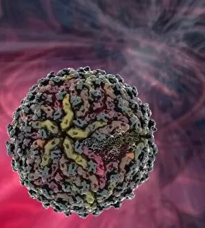
"Unveiling the World of Vector-Borne Diseases: A Microscopic Journey" Step into the microscopic realm and explore the intricate world of vector-borne diseases. From sleeping sickness parasites to malaria parasites, these tiny organisms hold immense power in their minuscule forms. In a transmission electron microscope (TEM), we witness the chilling sight of malaria parasites invading red blood cells. Their sinister presence reminds us of the devastating impact they have on millions around the globe. Dengue fever virus particles, also captured by TEM, reveal their spherical shape and intricate structure. These viral invaders are responsible for causing widespread outbreaks that leave communities vulnerable and in despair. Eastern equine encephalitis virus, another formidable foe, reveals its menacing appearance under TEM. Its complex architecture serves as a stark reminder of the neurological havoc it wreaks upon its unfortunate victims. Switching gears to scanning electron microscopy (SEM), we encounter sleeping sickness parasites up close and personal. Their elongated bodies with flagella protruding serve as a haunting visual representation of this deadly disease's parasitic nature. The SEM images continue to captivate our attention as multiple sleeping sickness parasites cluster together like an army ready for invasion. The sheer number is both awe-inspiring and terrifying at once, emphasizing their ability to spread rapidly through populations. Vesicular stomatitis virus takes center stage under TEM C016 / 4244 and TEM C016 / 4245 images. These spherical entities showcase their distinctive surface features while reminding us of the potential threat they pose to livestock and humans alike. Returning back to TEM imagery, we witness malaria parasites stealthily infiltrating red blood cells once again – a vivid depiction of how these cunning organisms manipulate our own bodies for survival. As we delve deeper into this microscopic journey through various imaging techniques, one thing becomes clear: vector-borne diseases are not just invisible threats but powerful adversaries capable of causing immense suffering worldwide.