
Viral Collection
"Unveiling the Intricate World of Viruses: From Coronavirus to Norovirus, Hepatitis C to Influenza" In this captivating illustration


All Professionally Made to Order for Quick Shipping
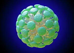
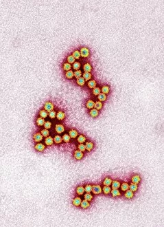
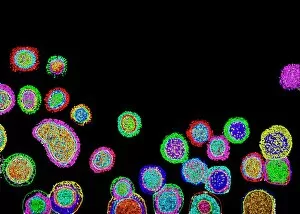
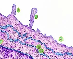
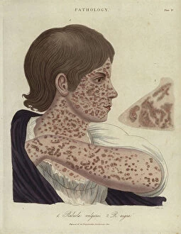
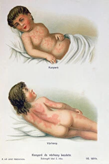
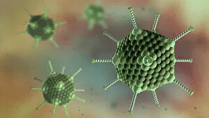
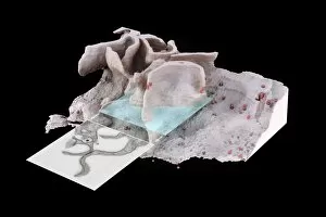
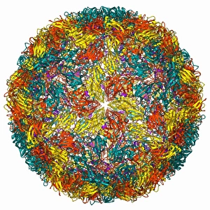
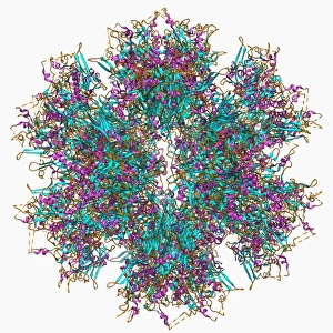
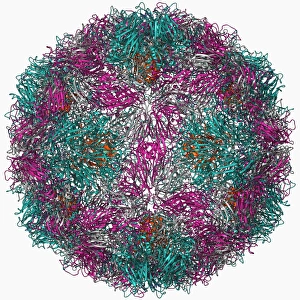
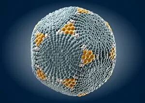
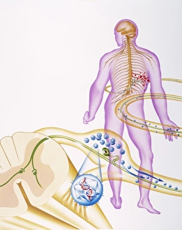
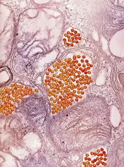
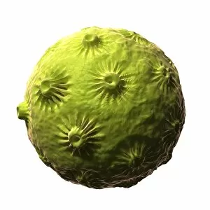
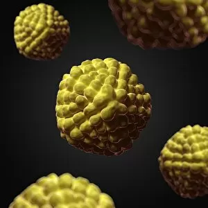
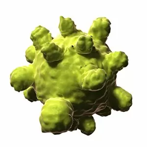
"Unveiling the Intricate World of Viruses: From Coronavirus to Norovirus, Hepatitis C to Influenza" In this captivating illustration, we delve into the microscopic realm of viruses that have left an indelible mark on our lives. Firstly, we encounter a mesmerizing depiction of the Coronavirus Structure Illustration. Its intricate spikes and spherical shape remind us of its global impact and ongoing battle against humanity. Moving forward, we witness Norovirus particles through a powerful TEM image. These tiny entities are notorious for causing stomach upsets and outbreaks in various settings. Next, our attention is drawn towards a molecular model showcasing the Hepatitis C virus enzyme. This visual representation highlights the complexity behind this viral infection that affects millions worldwide. As we explore further, an artistic rendering presents Flu virus particle artwork F008/3245. It serves as a reminder of how influenza can swiftly spread across populations during seasonal outbreaks. The dangers posed by infections transmitted through sneezing are vividly depicted in artwork C013/5949. The visualization captures the moment when pathogens disperse into the air with every forceful expulsion from an infected individual's respiratory system. Returning to coronavirus territory, another TEM image showcases Coronavirus particles in all their glory. Their distinctive crown-like appearance reinforces their notoriety as agents responsible for severe respiratory illnesses like SARS and MERS. Shifting gears slightly, an artistic portrayal introduces us to Adenovirus - known for causing respiratory infections but also harnessed as vectors in gene therapy research due to their unique properties. Our journey continues with yet another TEM image revealing Influenza virus particles - these minuscule culprits have been responsible for numerous pandemics throughout history and continue to pose significant health threats today. Stepping away momentarily from respiratory viruses, we observe Herpes virus replicating within cells under computer-generated artwork. This glimpse into its life cycle reminds us of the persistence and challenges associated with managing this widespread infection.