
Virion Collection
A virion is a fascinating microscopic entity that plays a crucial role in the world of viruses and infectious diseases


All Professionally Made to Order for Quick Shipping
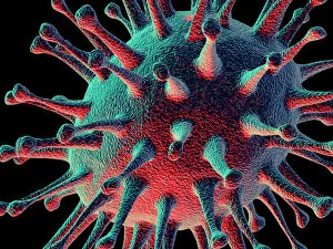
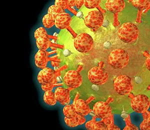
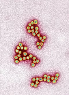
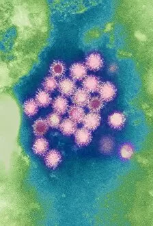
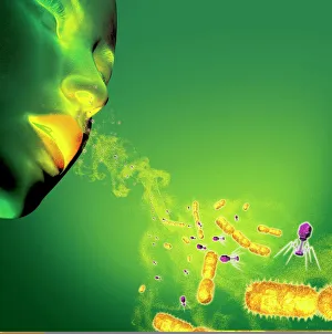
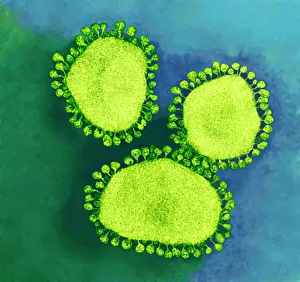
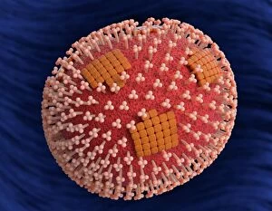
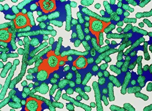
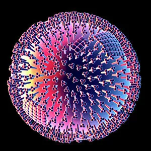
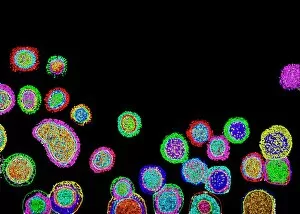
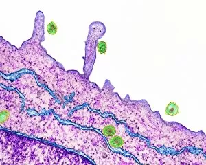
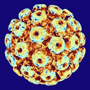
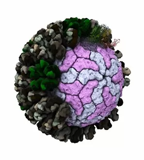
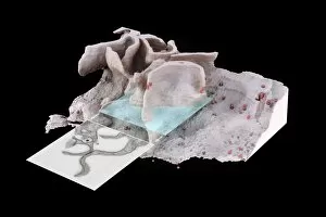
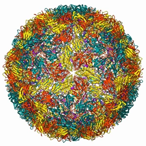
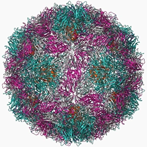
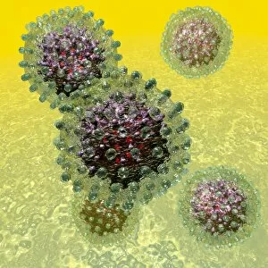
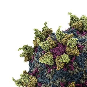
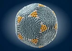
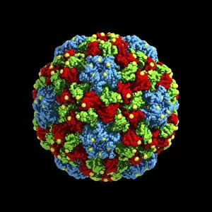
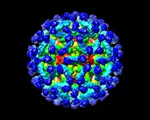
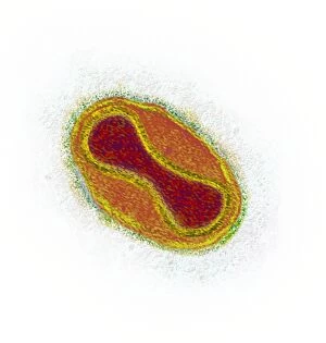
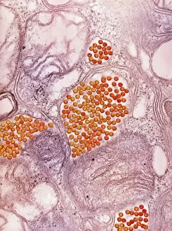
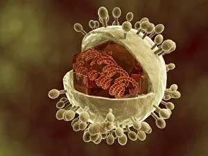
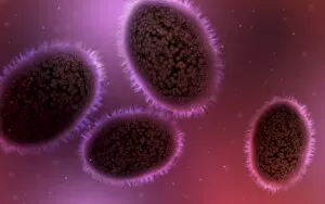
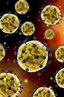
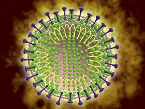
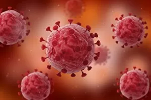
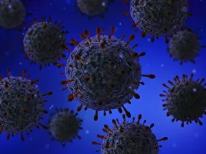
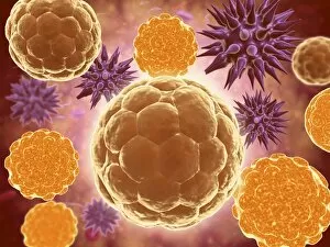
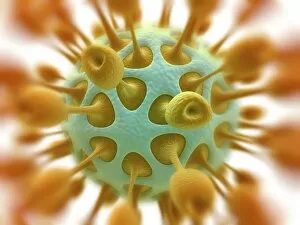
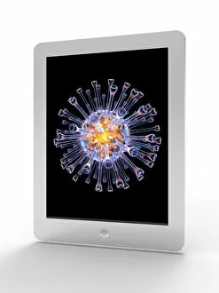
A virion is a fascinating microscopic entity that plays a crucial role in the world of viruses and infectious diseases. From the avian flu virus to HIV particles, norovirus particles, and hepatitis B viruses, these tiny structures are responsible for causing widespread infections. Under the powerful lens of a transmission electron microscope (TEM), we can observe the intricate details of various virions. The avian flu virus appears as an ominous presence with its spiky outer surface, while HIV particles reveal their complex structure that enables them to invade human immune cells. Norovirus particles captured by TEM showcase their round shape and distinctive pattern, reminding us of the havoc they wreak on our digestive system during outbreaks. Sneezing becomes more alarming when we realize it can spread infections like wildfire, as depicted in artwork C013 / 5949. Coronavirus particles also make an appearance under TEM; their crown-like spikes serve as a reminder of how this family of viruses has brought about global health crises such as SARS-CoV-2. Adenoviruses stand out with their geometric shapes portrayed in stunning artwork. Hepatitis B viruses take center stage once again due to their persistence and ability to cause chronic liver disease. Computer-generated artwork showcases influenza virus' unique structure while TEM images capture its spherical form along with other strains. Studying virions provides valuable insights into understanding infectious diseases and developing effective countermeasures against them. These captivating entities remind us of the constant battle between humans and pathogens at a microscopic level.