Fraud Blocker Skip to main content

Virology Collection

Virology, the fascinating study of viruses and their impact on living organisms

Background imageVirology Collection: Avian flu virus

Avian flu virus, computer artwork. A virus is a tiny pathogenic particle comprising genetic material enclosed in a protein coat. The coat contains surface proteins (spikes)

Background imageVirology Collection: Herpes virus replicating

Herpes virus replicating, computer artwork.Viruses are only able to replicate in a host cell.The glycoprotein spikes in the virus envelope(green)

Background imageVirology Collection: HIV particle

HIV particle, computer artwork. HIV (human immunodeficiency virus) causes AIDS (acquired immune deficiency syndrome). The virus consists of an RNA genome

Background imageVirology Collection: Avian flu virus

Avian flu virus, computer artwork. A virus is a tiny pathogenic particle comprising genetic material enclosed in a protein coat. The coat contains surface proteins (spikes)

Background imageVirology Collection: Norovirus particles, TEM

Norovirus particles, TEM
Norovirus particles. Coloured transmission electron micrograph (TEM) of norovirus particles. Norovirus is a genus of RNA (ribonucleic acid)

Background imageVirology Collection: HIV reverse transcription enzyme

HIV reverse transcription enzyme. Molecular models of the reverse transcriptase enzyme found in HIV (the human immunodeficiency virus)

Background imageVirology Collection: Hepatitis C virus enzyme, molecular model

Hepatitis C virus enzyme, molecular model
Hepatitis C virus enzyme. Molecular model of a genetic enzyme from the Hepatitis C virus. This enzyme is called HC-J4 RNA polymerase

Background imageVirology Collection: Hepatitis B viruses

Hepatitis B viruses
Hepatitis B virus. Coloured transmission electron micrograph (TEM) of hepatitis B viruses (circles), a cause of liver inflammation

Background imageVirology Collection: Microscopic view of human respiratory syncytial virus

Microscopic view of human respiratory syncytial virus (RSV). RSV causes respiratory tract infection of the lungs and breathing passages

Background imageVirology Collection: Infections spread by sneezing, artwork C013 / 5949

Infections spread by sneezing, artwork C013 / 5949
Infections spread by sneezing. Computer artwork showing virus particles (virions, blue) and bacteria (rod-shaped) in the spray produced by someone when they sneeze

Background imageVirology Collection: Coronavirus particles, TEM

Coronavirus particles, TEM
Coronavirus particles. Coloured transmission electron micrograph (TEM) of three coronavirus particles. Different strains of coronavirus are responsible for diseases such as the common cold

Background imageVirology Collection: Flu virus particle, artwork F008 / 3245

Flu virus particle, artwork F008 / 3245
Flu virus particle, computer artwork. In the virus envelope are two types of protein spike, haemagglutinin (H, pink) and neuraminidase (N, orange), which determine the strain of virus

Background imageVirology Collection: Hepatitis B viruses

Hepatitis B viruses

Background imageVirology Collection: Influenza virus, computer artwork

Influenza virus, computer artwork
Influenza virus. Computer artwork of an influenza (flu) virus. The virus consists of a core of RNA (ribonucleic acid) genetic material surrounded by a protein coat

Background imageVirology Collection: Influenza virus particles, TEM

Influenza virus particles, TEM
Influenza virus particles, coloured transmission electron micrograph (TEM). This virus is the cause of the respiratory disease influenza (flu) in humans and other animals

Background imageVirology Collection: Hepatitis C viruses, TEM

Hepatitis C viruses, TEM
Hepatitis C viruses. Coloured transmission electron micrograph (TEM) of hepatitis C virus particles (green) infecting cultured liver cells (purple)

Background imageVirology Collection: Adenovirus, artwork

Adenovirus, artwork
Adenovirus. Computer artwork of an adenovirus, showing the surface structure of the viruss outer protein coat (capsid). Adenoviruses are known to infect humans

Background imageVirology Collection: SARS coronavirus protein

SARS coronavirus protein. Molecular model of the ORF-9b protein produced by the SARS (severe acute respiratory syndrome) coronavirus

Background imageVirology Collection: Polyoma BK virus, artwork C013 / 7465

Polyoma BK virus, artwork C013 / 7465
Computer artwork of the capsid of a polyoma BK virus. This polyomavirus is common in the urinary tract of adults, where it lives without harming its host

Background imageVirology Collection: TEM of Lambda bacteriophages

TEM of Lambda bacteriophages

Background imageVirology Collection: Rift Valley fever virus, TEM

Rift Valley fever virus, TEM

Background imageVirology Collection: Paramyxovirus particles, TEM

Paramyxovirus particles, TEM
Sendai virus. Coloured transmission electron micrograph (TEM) of Sendai virus particles (virions, orange). The protein coat (capsid) of one of the particles has split

Background imageVirology Collection: Testing for viruses

Testing for viruses. A technician using a pipette to fill small sample vials. These are being used to test animal tissue samples for viruses such as avian influenza (bird flu) and swine fever

Background imageVirology Collection: Rotavirus particle, artwork

Rotavirus particle, artwork
Rotavirus particle. Cut-away artwork showing the structure of the rotavirus icosahedral capsid (protein coat). The capsid, which encloses the RNA (ribonucleic acid) genome

Background imageVirology Collection: Rhinovirus and antibody, molecular model C015 / 7139

Rhinovirus and antibody, molecular model C015 / 7139
Rhinovirus. Molecular model of the antigen-binding fragment (Fab) from a strongly neutralising antibody bound to a human rhinovirus 14 (HRV-14) particle

Background imageVirology Collection: Cold sore C014 / 4604

Cold sore C014 / 4604
Cold sore. Close-up of a cold sore beneath the nose of a 27-year-old female patient. Cold sores are caused by the Herpes simplex virus

Background imageVirology Collection: International biohazard symbol

International biohazard symbol warning of a potential biological hazard. This symbol is used worldwide in laboratories and on containers where biologically active agents are present

Background imageVirology Collection: Scanning electron micrograph of HIV particles infecting a human H9 T cell

Scanning electron micrograph of HIV particles infecting a human H9 T cell

Background imageVirology Collection: Stylized rabies virus particles

Stylized rabies virus particles, the cause of the viral neuroinvasive disease acute encephalitis

Background imageVirology Collection: Microscopic view of Neisseria gonorrhoeae

Microscopic view of Neisseria gonorrhoeae. Neisseria gonorrhoeae typically infects the mucous membranes causing infections such as urethritis, cervicitis, salpingitis, pelvic inflammatory disease

Background imageVirology Collection: Microscopic view of yellow fever virus

Microscopic view of yellow fever virus. Yellow fever is an acute viral disease

Background imageVirology Collection: Kaposis sarcoma on the skin of an AIDS patient

Kaposis sarcoma on the skin of an AIDS patient

Background imageVirology Collection: Conceptual image of rabies virus

Conceptual image of rabies virus

Background imageVirology Collection: HIV infected macrophage, SEM C018 / 8598

HIV infected macrophage, SEM C018 / 8598
HIV infected macrophage. Coloured ion-abrasion scanning electron micrograph (IA-SEM) of a macrophage white blood cell infected with human immunodeficiency virus (HIV, red)

Background imageVirology Collection: Foot-and-mouth disease virus F006 / 9556

Foot-and-mouth disease virus F006 / 9556
Foot-and-mouth disease virus. Molecular model of the foot-and-mouth disease (FMD) virus (Aphtae epizooticae) protein coat (capsid)

Background imageVirology Collection: Adenovirus penton base protein F006 / 9542

Adenovirus penton base protein F006 / 9542
Adenovirus penton base protein, molecular model. This protein molecule is a subunit called a penton, forming the vertices of the capsid of this adenovirus

Background imageVirology Collection: Rhinovirus 16 capsid, molecular model F006 / 9431

Rhinovirus 16 capsid, molecular model F006 / 9431
Rhinovirus 16 capsid, molecular model. This is human rhinovirus 16. The rhinovirus infects the upper respiratory tract and is the cause of the common cold. It is spread by coughs and sneezes

Background imageVirology Collection: Hepatitis B virus particles, artwork C016 / 9097

Hepatitis B virus particles, artwork C016 / 9097
Hepatitis B virus (HBV) particles, computer artwork. Each particle consists of an outer lipid envelope (green) that surrounds a protein nucleopcapsid (purple)

Background imageVirology Collection: Rhinovirus and antibody, molecular model C015 / 7138

Rhinovirus and antibody, molecular model C015 / 7138
Rhinovirus. Molecular model of the antigen-binding fragment (Fab) from a strongly neutralising antibody bound to a human rhinovirus 14 (HRV-14) particle

Background imageVirology Collection: Adenovirus hexon protein

Adenovirus hexon protein, molecular model. Hexon proteins are part of the protein coat or shell (capsid) of adenoviruses. In viruses

Background imageVirology Collection: Influenza virus particles, TEM

Influenza virus particles, TEM
Influenza virus particles, coloured transmission electron micrograph (TEM). The virus consists of ribonucleic acid (RNA), surrounded by a nucleocapsid (blue) and a lipid envelope (green)

Background imageVirology Collection: Parvovirus particle, artwork C013 / 4640

Parvovirus particle, artwork C013 / 4640
Parvovirus particle. Computer artwork showing the outer structure of a human parvovirus (family Parvoviridae) particle (virion)

Background imageVirology Collection: Cucumber mosaic virus, computer model

Cucumber mosaic virus, computer model
Cucumber mosaic virus (CMV), computer model. This image was created using molecular modelling software and data from X-ray crystallography

Background imageVirology Collection: HIV viruses, TEM

HIV viruses, TEM
HIV viruses. Coloured transmission electron micrograph (TEM) of human immunodeficiency virus (HIV) particles. These particles are docking at a T-lymphocyte (white blood cell) just before infection

Background imageVirology Collection: Murine norovirus with antibody fragments

Murine norovirus with antibody fragments
Murine norovirus (MNV) with antibody fragments, computer model. This image was created using molecular modelling software and data from cryo- electron microscopy

Background imageVirology Collection: AIDS virus

AIDS virus. Conceptual computer artwork of many HIV (human immunodeficiency virus) particles that are sectioned to show their internal structures

Background imageVirology Collection: Sindbis virus capsid protein

Sindbis virus capsid protein, molecular model

Background imageVirology Collection: Tobacco necrosis virus research, artwork

Tobacco necrosis virus research, artwork
Tobacco necrosis virus research, computer artwork


All Professionally Made to Order for Quick Shipping

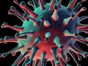
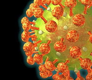
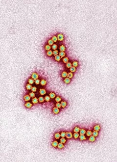
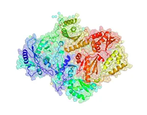
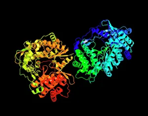
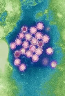
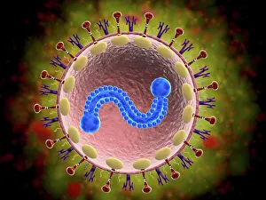
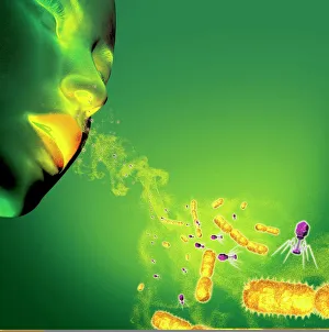
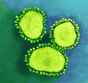
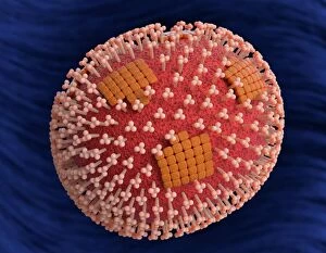
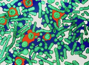
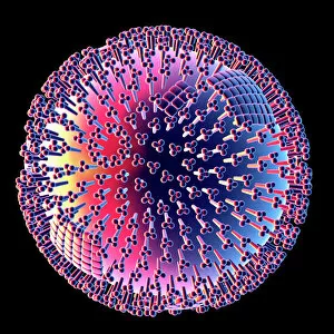
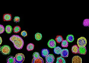
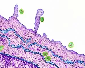
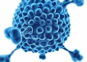
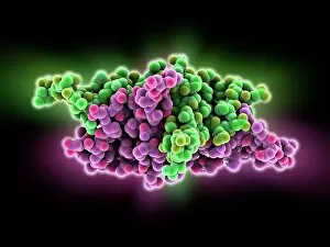
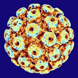
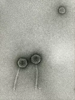
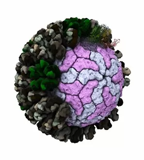
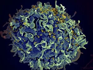
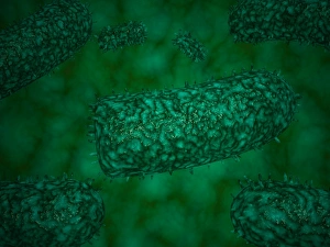
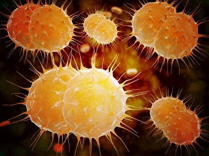
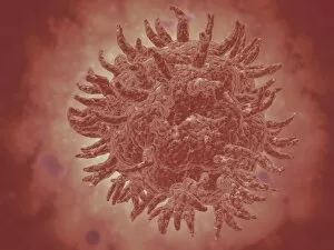
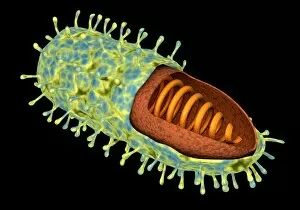
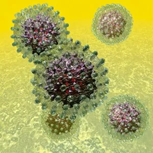
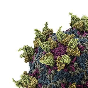
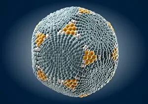
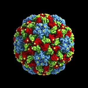
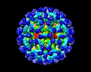
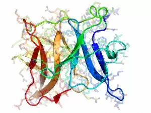
Virology, the fascinating study of viruses and their impact on living organisms, delves into a vast array of viral entities that have shaped our understanding of infectious diseases. From the Avian flu virus to HIV particles, Norovirus particles observed through TEM, and even the intricate workings of HIV reverse transcription enzyme and Hepatitis C virus enzyme molecular models - virologists explore these tiny but powerful agents with great curiosity. The Avian flu virus has long been a concern due to its potential for devastating outbreaks in both avian species and humans. Its ability to jump from birds to humans highlights the ever-present threat posed by zoonotic infections. Similarly, HIV particles continue to challenge researchers as they strive for effective treatments against this relentless retrovirus. Norovirus particles captured under transmission electron microscopy (TEM) reveal their unique structure responsible for causing gastroenteritis outbreaks worldwide. Understanding these minute details aids in developing strategies to combat their spread. Hepatitis B viruses are another focus within virology research due to their significant impact on liver health globally. Molecular models help visualize the complex mechanisms employed by these viruses during infection. Microscopic views of human respiratory syncytial virus remind us of how easily respiratory infections can spread among individuals through coughing or sneezing. Artwork depicting such scenarios serves as a reminder of the importance of hygiene practices in preventing disease transmission. Flu virus particle artwork showcases its distinct appearance while emphasizing its continuous evolution and ability to cause seasonal epidemics worldwide. This visual representation helps scientists comprehend its structure better when designing vaccines each year. Coronavirus particles imaged using TEM highlight recent concerns surrounding emerging viral threats like SARS-CoV-2, which causes COVID-19. The ongoing pandemic underscores the urgency for further virological studies aimed at combating future outbreaks effectively. Lastly, adenoviruses capture attention with artistic renditions showcasing their structural intricacies while reminding us that not all viruses are harmful.