Fraud Blocker Skip to main content

Home > Popular Themes > DNA

Cell structure

 : Cell structure
Sold 2
In Stock
We accept all major payment options so you can pay for the things you need in the way that is most convenient for you

Choose Your Product


Science Photo Library Photo Prints and Wall Art

Cell structure

Cell structure. Confocal light micrograph of cultured endothelial cells. A fluorescent dye has been used to show the cell structure. Nuclei (blue) contain the cells genetic information, DNA (deoxyribonucleic acid) packaged in chromosomes. Actin filaments part of the cell cytoskeleton are green. The cytoskeleton is responsible for the structure and motility of the cell. Actin is the most abundant cellular protein. The golgi apparatus, or body (red), is a membrane-bound organelle that modifies and packages proteins. It consists of long flattened vesicles arranged in stacks. Endothelial cells are flat and line all of the bodys blood vessels. Science Photo Library features Science and Medical images including photos and illustrations. © DAVID BECKER/SCIENCE PHOTO LIBRARY

Media ID 6400349

Actin Apparatus Bodies Body C Ulture Cellular Confocal Light Micrograph Confocal Light Microscope Cultured Cytology Cytoskeleton Dyes Edothelial Endothelia Endothelium F Actin Filament Filamentous Filaments Fluorescence Fluorescent Fluorescent Dye Golgi Immunofluorescence Immunofluorescent Immunostained Membrane Bound Motility Nuclei Nucleus Organelle Stained Structural Vascular Cells Protein

FEATURES IN THESE COLLECTIONS

> Europe > Italy > Basilicata > Venosa

> Popular Themes > DNA

SECURE PAYMENTS
We accept all major payment options so you can pay for the things you need in the way that is most convenient for you

Made in Australia
Safe Shipping, Easy Returns

FREE PERSONALISATION
We are proud to offer a range of customisation including Cropping, Captions, and Filters. Zoom Cropping can be adjusted in the basket


EDITORS COMMENTS

This print showcases the intricate structure of endothelial cells, as captured through a confocal light microscope. The fluorescent dye used in this image highlights various components of the cell, revealing its inner workings. The blue nuclei contain DNA, which carries the cell's genetic information and is packaged in chromosomes. Green actin filaments form part of the cell cytoskeleton, responsible for maintaining its structure and enabling movement. Actin is a vital protein found abundantly within cells. The red-colored golgi apparatus stands out prominently in this image; it is a membrane-bound organelle that plays a crucial role in modifying and packaging proteins within the cell. Composed of long flattened vesicles arranged in stacks, it ensures efficient protein transport. Endothelial cells are flat and line all blood vessels throughout our bodies. They play an essential role in vascular biology by regulating blood flow and vessel permeability. This stunning micrograph not only provides valuable insights into cellular structures but also serves as a testament to the remarkable complexity present within even seemingly simple organisms like individual cells. It reminds us of the beauty hidden beneath our skin – an intricate world that fuels life itself.


Framed Prints of Cell structure

Discover the intricacies of life with our Media Storehouse Framed Prints featuring the captivating image of "Cell Structure" by Science Photo Library. This confocal light micrograph showcases the mesmerizing beauty of cultured endothelial cells, illuminated by the use of a fluorescent dye. Delve deeper into the world of biology and bring the wonders of science into your home or office with our high-quality framed prints. Each print is meticulously crafted with attention to detail, ensuring a stunning display that is sure to impress. Order yours today and ignite your curiosity!


Photo Prints of Cell structure

Discover the intricacies of life with Media Storehouse's Scientific Photographic Prints. Feast your eyes on this captivating image of a confocal light micrograph, courtesy of Science Photo Library. Witness the mesmerizing beauty of cultured endothelial cells as they come to life in vibrant colors. The intricate details of the cell structure are brought to the forefront, revealing a world that is both complex and awe-inspiring. Perfect for scientific institutions, laboratories, or any space that appreciates the wonders of the natural world. Add this print to your collection and ignite the curiosity within.


Poster Prints of Cell structure

"Discover the intricacies of cellular biology with our Media Storehouse Poster Prints featuring the stunning image, "Cell Structure" from Science Photo Library. This captivating confocal light micrograph showcases the complex beauty of cultured endothelial cells, highlighted through the use of a fluorescent dye. Bring this fascinating glimpse into the microscopic world into your home or office, and ignite curiosity and inspiration with every glance. Our high-quality prints deliver vibrant colors and sharp details, ensuring a stunning display that will captivate any audience."


Jigsaw Puzzles of Cell structure

Discover the intricacies of cellular biology with our captivating Media Storehouse Jigsaw Puzzles. This scientific masterpiece, featuring a confocal light micrograph of cultured endothelial cells from Science Photo Library, invites you on a challenging yet rewarding journey. Observe the intricate details of cell structures as fluorescent dyes highlight various components. Engage your mind and enhance your understanding of science, one piece at a time.

diamond
Made With High Grade Materials
thumb_up
Professional Quality Finish
package_2
Carefully Packed To Aid Safe Arrival

You May Also Like


Glial cellsGlial cells
Add to Basket
Bubbles of Black Dry Stout (LM) C014 / 1712Bubbles of Black Dry Stout (LM) C014 / 1712
Add to Basket
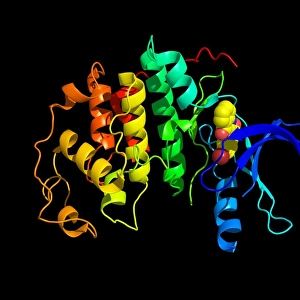
Kinase molecule, secondary structureKinase molecule, secondary structure
Add to Basket
Induced nerve stem cells, micrographInduced nerve stem cells, micrograph
Add to Basket
Nereids and Triton, c1753-1762, (1928). Artist: Giovanni Battista TiepoloNereids and Triton, c1753-1762, (1928). Artist: Giovanni Battista Tiepolo
Add to Basket
PARIS-SEINE-SUMMERPARIS-SEINE-SUMMER
Add to Basket
spa1962 bw neg pub 008 edited-1spa1962 bw neg pub 008 edited-1
Add to Basket
Dave Pither (Matchless) 1987 Classic Manx Grand PrixDave Pither (Matchless) 1987 Classic Manx Grand Prix
Add to Basket
Annunciation Basilica door sculpture depicting Adam and Eve, Nazareth, Galilee, IsraelAnnunciation Basilica door sculpture depicting Adam and Eve, Nazareth, Galilee, Israel
Add to Basket
Elevation overlooking the 10th tee, West Course, Winged Foot Golf Club, Mamaroneck, New York, 1925Elevation overlooking the 10th tee, West Course, Winged Foot Golf Club, Mamaroneck, New York, 1925
Add to Basket
The Mars and L Hercule, c1799. Artist: Nicholas PocockThe Mars and L Hercule, c1799. Artist: Nicholas Pocock
Add to Basket
Paper making, 1751-1777Paper making, 1751-1777
Add to Basket

Gallery Wall Inspiration

Watson and Crick with their DNA model
Rosalind Franklin
DNA molecule
DNA transcription, molecular model
Double-stranded RNA molecule
DNA molecule
Watson and Crick, DNA discovers
DNA molecule, artwork
The Tarbet Hotel, Loch Lomond, Caledonian Railway poster, 1920
Peebles for Health, Rest and Recreation, railway poster, 1900-1922
Computer screen showing a human genetic sequence
X and Y chromosomes
DNA molecule
Mitosis, light micrograph
Rough endoplasmic reticulum, TEM
Cell structure
Herpes virus replicating
Fingerprint scanner
Computer artwork of a beta DNA segment and spheres
Nucleotide base matrix
DNA molecule, computer model
Nucleosome molecule
DNA molecule, abstract image
DNA nucleosome, molecular model
Dividing yeast cells, SEM
E. coli bacterium


© Copyright Media Storehouse, All Rights Reserved 2003 - 2025
Facebook     Pinterest     100% Payment Secure     Reviews IO