Home > Popular Themes > Human Body
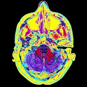
Coloured illustration of sectioned brain in head

![]()
Wall Art and Photo Gifts from Science Photo Library
Coloured illustration of sectioned brain in head
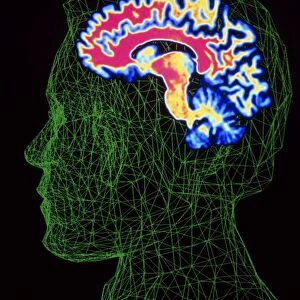
Brain. Computer-coloured illustration of a human head in side view, sectioned to show the brain and its blood vessels. The face is at right; cavities (pink) of the nose, mouth and pharynx are seen. Cerebral blood vessels (at top) branch within the cerebrum (yellow) of the brain. The large cerebrum of the brain is responsible for memory, conscious thought and voluntary movement. The cerebellum is a tree-shaped structure (blue/pink) at ear level. At the top of the yellow spinal cord is a swelling that forms the brainstem responsible for primitive reflexes. Illustration by the Italian anatomist Paolo Mascagni (1755-1815), author of the famous treatise: " Vasorum Lymphaticorum Corporis Humani"
Science Photo Library features Science and Medical images including photos and illustrations
Media ID 6421558
© MEHAU KULYK/SCIENCE PHOTO LIBRARY
Blood System Brain Anatomy Brain Stem Central Nervous System Cerebellum Cerebrum Mascagni Nasal Cavity Brain
FEATURES IN THESE COLLECTIONS
EDITORS COMMENTS
This print showcases a computer-coloured illustration of a sectioned human brain within the confines of a head. The intricate details reveal the complexity and beauty of our most vital organ. At first glance, one's attention is drawn to the face on the right side, while pink cavities representing the nose, mouth, and pharynx are visible. The focal point lies in the yellow cerebrum, which dominates this artistic representation. This large structure plays a crucial role in memory retention, conscious thought processes, and voluntary movement control. Branching out from above are cerebral blood vessels that intricately weave their way through this essential part of our brain. A striking feature is the tree-shaped cerebellum depicted in shades of blue and pink at ear level. Positioned at the top of the spinal cord is a swelling forming what is known as the brainstem—a key component responsible for primitive reflexes. This remarkable artwork was created by Paolo Mascagni (1755-1815), an esteemed Italian anatomist renowned for his treatise titled "Vasorum Lymphaticorum Corporis Humani". His meticulous attention to detail shines through in this illustration that provides viewers with an insightful glimpse into our central nervous system's inner workings. Science Photo Library has masterfully captured this image—an amalgamation of artistry and anatomy—showcasing its profound significance within medical science and research.
MADE IN AUSTRALIA
Safe Shipping with 30 Day Money Back Guarantee
FREE PERSONALISATION*
We are proud to offer a range of customisation features including Personalised Captions, Color Filters and Picture Zoom Tools
SECURE PAYMENTS
We happily accept a wide range of payment options so you can pay for the things you need in the way that is most convenient for you
* Options may vary by product and licensing agreement. Zoomed Pictures can be adjusted in the Cart.