Home > Science > SEM
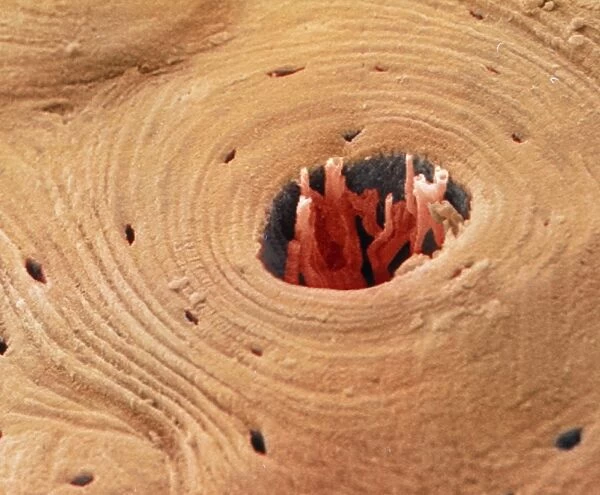
Coloured SEM of transverse section of compact bone
![]()

Wall Art and Photo Gifts from Science Photo Library
Coloured SEM of transverse section of compact bone
Compact bone. Coloured scanning electronmicrograph (SEM) of a transverse section ofcompact bone. The Haversian canal (dark centralarea) contains blood vessels, lymphatics andnerves (all coloured red). Haversian canals areinterconnecting neurovascular channels which runparallel to the long axes of long bones.Concentric bony layers, the lamellae, enclose theHaversian canals. The lamellae are made ofcompacted collagen fibres and ground substancesproduced by specialised cells called osteoblasts.During bone formation the osteoblasts becomeprogressively trapped in lacunae (small darkareas). Magnification: x315 at 6x7cm size. x740 at8x6", x395 at 10x7cm master size
Science Photo Library features Science and Medical images including photos and illustrations
Media ID 9303939
© POWER AND SYRED/SCIENCE PHOTO LIBRARY
Bones Circle Circles Compact Compact Bone Haversian Canal Lacuna Lamella Magnified Image Microscopic Photos Round Shape Rounded Circular Subjects
FEATURES IN THESE COLLECTIONS
EDITORS COMMENTS
This print showcases a coloured scanning electron micrograph (SEM) of a transverse section of compact bone. The intricate details are brought to life as the Haversian canal, depicted in a dark central area, reveals its vital contents: blood vessels, lymphatics, and nerves all beautifully coloured red. These Haversian canals serve as interconnected neurovascular channels that run parallel to the long axes of long bones. Enclosing these essential canals are concentric bony layers known as lamellae. Composed of compacted collagen fibers and ground substances produced by specialized cells called osteoblasts, the lamellae play a crucial role in maintaining bone strength and structure. The process of bone formation is visually represented through the progressively trapped osteoblasts found within small dark areas known as lacunae. This magnified image allows us to appreciate their significance in building and maintaining healthy bones. With a magnification level reaching x315 at 6x7cm size, x740 at 8x6", and x395 at 10x7cm master size, this microscopic photograph captures the complexity and beauty hidden within our skeletal system. It serves as an awe-inspiring reminder of the remarkable intricacy present within our own bodies.
MADE IN AUSTRALIA
Safe Shipping with 30 Day Money Back Guarantee
FREE PERSONALISATION*
We are proud to offer a range of customisation features including Personalised Captions, Color Filters and Picture Zoom Tools
SECURE PAYMENTS
We happily accept a wide range of payment options so you can pay for the things you need in the way that is most convenient for you
* Options may vary by product and licensing agreement. Zoomed Pictures can be adjusted in the Cart.


