Home > Popular Themes > Human Body
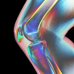
Coloured X-ray of a human knee joint (front view)
")
![]()
Wall Art and Photo Gifts from Science Photo Library
Coloured X-ray of a human knee joint (front view)
Knee joint. Coloured X-ray of a healthy human knee joint, in front view. Two bones meet at the knee, forming a joint that works like a hinge. At top is the large femur (thigh-bone), which articulates with the tibia (shin-bone) at bottom. Next to the tibia (at lower right) is the smaller fibula bone. The patella or kneecap (above centre, blue) is a protective bone at the front of the knee held in position by muscles and tendons. Two discs of protective cartilage cover the surfaces of the femur and tibia to reduce friction between these bones. This joint, the largest in the body, allows a backward-forward hinge movement with slight rotation
Science Photo Library features Science and Medical images including photos and illustrations
Media ID 6419956
© MEHAU KULYK/SCIENCE PHOTO LIBRARY
Bones Diagnosis Femur Joint Knee Knee Cap Knee Joint Patella Technique Tibia Hinge Joint
EDITORS COMMENTS
This print showcases a coloured X-ray of a human knee joint, providing an intricate glimpse into the inner workings of our bodies. The front view reveals the remarkable complexity and harmony between bones, muscles, tendons, and cartilage that enable us to move with ease. At the center of attention is the knee joint itself – a hinge-like structure where two crucial bones meet. The mighty femur, or thigh-bone, dominates the top portion while elegantly articulating with its counterpart below - the tibia or shin-bone. Adjacent to the tibia rests its smaller companion, known as fibula bone. A striking blue hue draws our eyes towards another key component: the patella or kneecap. Positioned above center stage, this protective bone owes its stability to surrounding muscles and tendons. To ensure smooth movement within this vital joint, two discs of protective cartilage gracefully cover both surfaces of the femur and tibia. Their presence significantly reduces friction between these weight-bearing bones. Undoubtedly one of nature's marvels, this largest joint in our body allows for backward-forward hinge movements accompanied by slight rotation when needed. This mesmerizing image not only unveils astonishing anatomical details but also serves as a valuable tool for medical professionals in diagnosing various conditions related to this intricate region. Through science and technology merging seamlessly in this photograph from Science Photo Library, we gain deeper insights into our own physicality – a testament to both artistry and scientific discovery.
MADE IN AUSTRALIA
Safe Shipping with 30 Day Money Back Guarantee
FREE PERSONALISATION*
We are proud to offer a range of customisation features including Personalised Captions, Color Filters and Picture Zoom Tools
SECURE PAYMENTS
We happily accept a wide range of payment options so you can pay for the things you need in the way that is most convenient for you
* Options may vary by product and licensing agreement. Zoomed Pictures can be adjusted in the Cart.