Fraud Blocker Skip to main content

Home > Science > SEM

Dust mites

 : Dust mites
In Stock
We accept all major payment options so you can pay for the things you need in the way that is most convenient for you

Choose Your Product

Framed Photos
Buy from AU$119.99
Photo Prints
Buy from AU$12.99
Jigsaw Puzzles
Buy from AU$89.99
Poster Prints
Buy from AU$24.99
Canvas Prints
Buy from AU$124.99
Fine Art Prints
Buy from AU$92.53
Metal Prints
Buy from AU$43.99
Cards
Buy from AU$12.99
Home Decor
Buy from AU$24.99

Science Photo Library Photo Prints and Wall Art

Dust mites

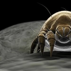
Dust mites. Coloured scanning electron micrograph (SEM) of two dust mites (Dermatophagoides sp.) on fabric fibres. Millions of dust mites inhabit the home, feeding on shed skin cells. They mainly live in furniture, and are invisible to the naked eye due to their size. The excrement and dead bodies of these mites may cause allergic reactions in susceptible people. Magnification: x150 at 6x7cm size. Science Photo Library features Science and Medical images including photos and illustrations. © POWER AND SYRED/SCIENCE PHOTO LIBRARY

Media ID 9331297

Allergen Allergic Reaction Arachnida Couple Fabric Fibres Household Material

FEATURES IN THESE COLLECTIONS

> Animals > Insects > Mites > Related Images

> Science > SEM

SECURE PAYMENTS
We accept all major payment options so you can pay for the things you need in the way that is most convenient for you

Made in Australia
Safe Shipping, Easy Returns

FREE PERSONALISATION
We are proud to offer a range of customisation including Cropping, Captions, and Filters. Zoom Cropping can be adjusted in the basket


EDITORS COMMENTS

This print from Science Photo Library reveals the hidden world of dust mites, showcasing their intricate features and remarkable lifestyle. In this coloured scanning electron micrograph (SEM), two dust mites belonging to the Dermatophagoides species can be seen clinging onto fabric fibres. These minuscule creatures are a common presence in our homes, with millions of them thriving on shed skin cells. Despite their tiny size, these arachnids play a significant role in our daily lives. Living predominantly in furniture, they remain invisible to the naked eye but leave behind traces of their existence that can have profound effects on human health. The excrement and deceased bodies of these mites act as allergens, triggering allergic reactions in susceptible individuals. At a magnification level of x150 within a 6x7cm frame, this image offers an extraordinary glimpse into the microscopic world that coexists with us every day. It serves as a reminder that even though we may not see them directly, these seemingly insignificant creatures have an impact on our well-being. Through this photograph's vivid colors and precise detailing captured by SEM technology, it becomes evident how interconnected wildlife is with our household environment. This mesmerizing snapshot invites contemplation about the delicate balance between humans and nature while highlighting the unseen forces at work within our own homes.


Framed Prints of Dust mites

Discover the unseen world with our Media Storehouse Framed Prints featuring the captivating image of Dust Mites by Science Photo Library. This coloured scanning electron micrograph (SEM) reveals the intricate details of two dust mites (Dermatophagoides sp.) in their natural habitat on fabric fibers. A fascinating addition to any home or office, these framed prints showcase the wonders of science and the hidden world around us. Bring this microscopic marvel into your living space and ignite curiosity and conversation.


Photo Prints of Dust mites

Discover the unseen world with Media Storehouse's Photographic Prints. This captivating image, sourced from Science Photo Library, showcases a coloured scanning electron micrograph (SEM) of two dust mites (Dermatophagoides sp.) in their natural habitat – on fabric fibers. With millions of these microscopic creatures residing in our homes, feeding on shed skin cells, this print brings a fascinating perspective to the common dust mite. Bring this intriguing piece of scientific art into your space and ignite conversations.


Poster Prints of Dust mites

Discover the unseen world with Media Storehouse's captivating Poster Prints. This particular piece features a coloured scanning electron micrograph (SEM) of two dust mites (Dermatophagoides sp.) in their natural habitat on fabric fibers. With millions of these microscopic creatures residing in our homes, feeding on shed skin cells, this print is a fascinating reminder of the hidden world around us. Bring the wonders of science into your living space with Media Storehouse's high-quality, vibrant Poster Prints.


Jigsaw Puzzles of Dust mites

Discover the microscopic wonders of your home with Media Storehouse's captivating Jigsaw Puzzles. This intriguing puzzle, featuring a coloured scanning electron micrograph (SEM) of dust mites (Dermatophagoides sp.) by Science Photo Library, invites you on a mesmerizing journey into the unseen world. Millions of these tiny creatures inhabit our homes, feeding on shed skin cells. Assemble this puzzle to bring this fascinating image to life and unveil the hidden inhabitants of your living space. Engage your mind, stimulate your creativity, and deepen your appreciation for the world around you with Media Storehouse's Jigsaw Puzzles.

diamond
Made With High Grade Materials
thumb_up
Professional Quality Finish
package_2
Carefully Packed To Aid Safe Arrival

You May Also Like


Carpet beetleCarpet beetle
Add to Basket
SEM of a dust miteSEM of a dust mite
Add to Basket
Microscopic visualization of a dust miteMicroscopic visualization of a dust mite
Add to Basket

Gallery Wall Inspiration

Nerve cell, SEM
Red-barbed ant, SEM
Neutrophil engulfing MRSA, SEM C018 / 8596
Diatom alga, SEM
Diatom, SEM
X and Y chromosomes
Lavender pollen grain, SEM
Scanning Electron Micrograph (SEM): Tardigrade or ‘Water Bear Magnification x 1250 (A4 size: 29. 7 cm width)
Cat tongue surface, SEM
False-coloured SEM (Scanning Electron Micrograph) of a Tardigrade (Tardigrada) amongst moss, from Peak District National Park, Derbyshire, UK. This tardigrade is approximately 0. 1 millimetres long. Digital composite
Collagen, Scanning electron micrograph (SEM)
Santos-Dumont / Sem
T lymphocytes and cancer cell, SEM C001 / 1679
Tardigrade or Water Bear
Hazel pollen grains
Crysotile asbestos
Uterus lining during menstruation, SEM
Iris of the eye, SEM
Stylus in LP record
Salmonella bacteria, SEM
SEM of penicillin fungus
Vascular bundle, SEM
Geranium anther, SEM
Dahlia flower pollen, SEM
Scanning Electron Micrograph (SEM): Praying Mantis - Magnification x 30 (if print A4 size: 29. 7 cm wide)
Tardigrade, SEM
Scanning electron micrograph (SEM) of red blood cell


© Copyright Media Storehouse, All Rights Reserved 2003 - 2025
Facebook     Pinterest     100% Payment Secure     Reviews IO