Home > Science > SEM
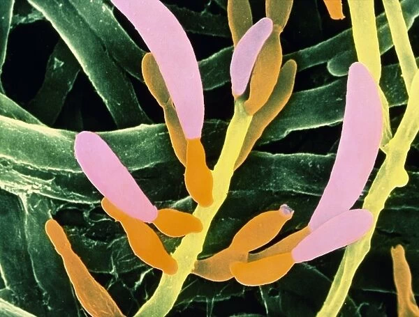
False-colour SEM of the fungus Fusarium oxysporum
![]()

Wall Art and Photo Gifts from Science Photo Library
False-colour SEM of the fungus Fusarium oxysporum
False-colour scanning electron micrograph of the pathogenic fungus Fusarium oxysporum, which causes wilt disease in tomato and carnation plants. The micrograph shows the hyphae, or vegetative structure of the fungus in the background coloured green. The pink and organge structures, branching off from main hyphal filaments, are conidiophores or specialised spore producing bodies. The conidia, or spores, are produced from the tips of these structures and when ripe are released into the soil. One such structure that has released its conidia is visible at top right showing a jagged crown-like opening. Magnification: x2000 at 6x4.4cm size
Science Photo Library features Science and Medical images including photos and illustrations
Media ID 6291961
© DR JEREMY BURGESS/SCIENCE PHOTO LIBRARY
Conidiophore Eumycota Fungal Fungi Fungus Mycology Naturemycology Pathogenic Plant Disease Type False Coloured
EDITORS COMMENTS
This print showcases a false-colour scanning electron micrograph of the pathogenic fungus Fusarium oxysporum. Known for causing wilt disease in tomato and carnation plants, this fungus is brought to life through vibrant hues and intricate details. The background is adorned with green-colored hyphae, representing the vegetative structure of the fungus. Branching off from these main hyphal filaments are striking pink and orange structures called conidiophores. These specialized spore producing bodies play a crucial role in the life cycle of the fungus. At their tips, they produce conidia or spores that are eventually released into the soil when ripe. A remarkable feature captured in this image is a conidiophore that has already discharged its conidia. Its jagged crown-like opening at the top right corner serves as evidence of this process taking place. The magnification level of x2000 allows us to appreciate even the tiniest intricacies within this microscopic world. As we delve into nature's wonders, it becomes evident how interconnected all living organisms truly are. This photograph not only highlights the beauty found within fungi but also sheds light on their impact on plant health and agriculture as pathogens like Fusarium oxysporum can cause devastating diseases. With its scientific significance and artistic allure, this print by Science Photo Library invites us to explore an unseen realm where nature's complexity unfolds before our eyes.
MADE IN AUSTRALIA
Safe Shipping with 30 Day Money Back Guarantee
FREE PERSONALISATION*
We are proud to offer a range of customisation features including Personalised Captions, Color Filters and Picture Zoom Tools
SECURE PAYMENTS
We happily accept a wide range of payment options so you can pay for the things you need in the way that is most convenient for you
* Options may vary by product and licensing agreement. Zoomed Pictures can be adjusted in the Cart.


