Home > Popular Themes > Human Body
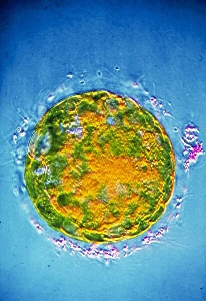
False-colour TEM of 6-day-old fertilised ovum
![]()

Wall Art and Photo Gifts from Science Photo Library
False-colour TEM of 6-day-old fertilised ovum
False-colour transmission electron micrograph (TEM) of the human morula - a fertilised ovum - 6 days after fertilisation. The morula is an early stage of embryonic development; it is formed by successive cleavage of cells in the fertilised ovum. It consists of a solid ball of cells & may be considered as an intermediate stage between the zygote, the fertilised ovum before the advent of cleavage, & the blastocyst, the hollow ball of cells with an inner mass that attaches itself to the wall of the uterus. The ring of material (pink) around the edge of the morula is debris of sperm that failed to penetrate & fertilise the ovum. Magnification: x35 at 35mm size
Science Photo Library features Science and Medical images including photos and illustrations
Media ID 6454759
© CNRI/SCIENCE PHOTO LIBRARY
EDITORS COMMENTS
This print showcases a false-colour transmission electron micrograph (TEM) of a 6-day-old fertilised ovum, specifically the human morula. The morula represents an early stage in embryonic development, formed through successive cleavage of cells within the fertilised ovum. It appears as a solid ball composed of numerous cells and serves as an intermediate phase between the zygote (the unfertilised ovum) and the blastocyst (a hollow ball with an inner mass that attaches to the uterine wall). The mesmerizing image reveals intricate details at a magnification of x35 in a 35mm size. The vibrant pink ring encircling the morula's edge signifies debris from sperm that failed to penetrate and fertilise the ovum, adding an intriguing element to this visual representation. Science Photo Library has expertly captured this momentous event in human reproduction, offering us a glimpse into the complex world of cellular activity during early embryogenesis. This photograph not only highlights scientific advancements but also evokes wonder and appreciation for life's beginnings.
MADE IN AUSTRALIA
Safe Shipping with 30 Day Money Back Guarantee
FREE PERSONALISATION*
We are proud to offer a range of customisation features including Personalised Captions, Color Filters and Picture Zoom Tools
SECURE PAYMENTS
We happily accept a wide range of payment options so you can pay for the things you need in the way that is most convenient for you
* Options may vary by product and licensing agreement. Zoomed Pictures can be adjusted in the Cart.


