Fraud Blocker Skip to main content

Home > Popular Themes > Human Body

Illustration of the human placenta

 : Illustration of the human placenta
Sold 4
In Stock
We accept all major payment options so you can pay for the things you need in the way that is most convenient for you

Choose Your Product


Science Photo Library Photo Prints and Wall Art

Illustration of the human placenta

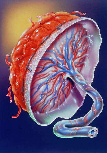
Illustration of the human placenta. The placenta is an organ that develops in the female uterus during pregnancy. Through it, and an umbilical cord (at lower right), the blood supply of the mother is linked to the fetus. A rich supply of placental blood vessels (red and blue, at centre) allow for nutrients and oxygen to be transferred from mother to fetus. And it allows waste products from the fetus to pass back to the mother. Apart from being a vital link between mother and fetus, the placenta also produces hormones. By the end of pregnancy it has grown about 20cm wide and 2.5cm thick. Shortly after birth, the placenta is expelled from the uterus as the afterbirth. Science Photo Library features Science and Medical images including photos and illustrations. © JOHN BAVOSI/SCIENCE PHOTO LIBRARY

Media ID 6422914

Placenta Re Production Reproductive System Umbilical Cord Female Anatomy

FEATURES IN THESE COLLECTIONS

> Animals > Mammals > Muridae > Grandis

> Animals > Mammals > Sciuridae > Grandis

> Popular Themes > Human Body

SECURE PAYMENTS
We accept all major payment options so you can pay for the things you need in the way that is most convenient for you

Made in Australia
Safe Shipping, Easy Returns

FREE PERSONALISATION
We are proud to offer a range of customisation including Cropping, Captions, and Filters. Zoom Cropping can be adjusted in the basket


EDITORS COMMENTS

This print beautifully captures the intricate illustration of the human placenta, a vital organ that develops within the female uterus during pregnancy. Positioned at the lower right corner is the umbilical cord, which serves as a lifeline connecting the mother's blood supply to that of her developing fetus. The central focus lies on an elaborate network of placental blood vessels depicted in shades of red and blue, symbolizing their crucial role in facilitating nutrient and oxygen transfer from mother to baby. The placenta also acts as a remarkable filter, enabling waste products generated by the growing fetus to be efficiently eliminated back into the mother's bloodstream. Beyond its pivotal function as a conduit between mother and child, this incredible organ is responsible for producing essential hormones throughout pregnancy. Measuring approximately 20cm wide and 2.5cm thick by full term, this awe-inspiring artwork showcases both its size and complexity. Following childbirth, when it has fulfilled its purpose, the placenta is naturally expelled from the uterus as part of what is commonly referred to as "the afterbirth". This stunning reproduction not only highlights key elements of female anatomy but also emphasizes how intricately connected our reproductive systems are with new life formation. Science Photo Library has masterfully captured this profound moment in human development through their exceptional artistry and attention to detail.


Framed Prints of Illustration of the human placenta

Introducing the Media Storehouse Framed Prints collection, featuring this captivating illustration of the human placenta from Science Photo Library. This stunning image offers a unique glimpse into the marvels of human biology. The placenta, an essential organ that develops in the uterus during pregnancy, is depicted here with an umbilical cord at the lower right. Our high-quality framed prints bring this intricate illustration to life, making it an excellent addition to any home or office space. Delve into the wonders of the human body and celebrate the miracle of life with this exquisite piece.


Photo Prints of Illustration of the human placenta

Discover the wonders of human biology with Media Storehouse's collection of Scientific Photographic Prints. This captivating illustration showcases the intricate structure of the human placenta, an essential organ that develops in the uterus during pregnancy. Through the placenta, vital nutrients and oxygen are transferred from the mother to the growing fetus via the umbilical cord. Bring a touch of scientific discovery into your home or office with this beautifully printed and framed piece. Perfect for educators, healthcare professionals, or anyone with a curiosity for the marvels of human anatomy.


Poster Prints of Illustration of the human placenta

Introducing the Media Storehouse range of Poster Prints featuring the captivating illustration of the human placenta. This scientific masterpiece, sourced from Science Photo Library, offers a unique and educational insight into the marvels of human biology. The placenta, an essential organ that develops in the uterus during pregnancy, is beautifully illustrated here with an umbilical cord attached at the lower right. Bring this intriguing print into your home or office to inspire curiosity and foster a deeper understanding of the intricacies of life. Order now and experience the wonders of science in high-quality print form.


Jigsaw Puzzles of Illustration of the human placenta

Discover the wonders of human anatomy with our captivating jigsaw puzzles from Media Storehouse. This intriguing puzzle showcases an illustration of the human placenta, an essential organ that develops in the uterus during pregnancy. Assemble the pieces to reveal the intricate details of this remarkable organ, through which essential nutrients and oxygen are transferred from the mother to the growing fetus via the umbilical cord. Ideal for science enthusiasts, educators, and anyone interested in the mysteries of life, this puzzle promises a stimulating and rewarding experience.

diamond
Made With High Grade Materials
thumb_up
Professional Quality Finish
package_2
Carefully Packed To Aid Safe Arrival

You May Also Like


Female body showing digestive and circulatory systemFemale body showing digestive and circulatory system
Add to Basket
X-ray view of a woman sitting on floor with skeletal bones superimposedX-ray view of a woman sitting on floor with skeletal bones superimposed
Add to Basket
1st Team Player Images 2007-81st Team Player Images 2007-8
Add to Basket
Greyhound TrainingGreyhound Training
Add to Basket
Wimbledon Tea IntervalWimbledon Tea Interval
Add to Basket
Dog - Basset Hound wearing hat in vanDog - Basset Hound wearing hat in van
Add to Basket
Woodland K991356Woodland K991356
Add to Basket
Rangers Football Club: 1896-1897 Champions - Triple Cup Victory SquadRangers Football Club: 1896-1897 Champions - Triple Cup Victory Squad
Add to Basket
Arsenal's Victory: Emirates Stadium, 2-0 Blackburn Rovers, Barclays Premier League, 11/2/08Arsenal's Victory: Emirates Stadium, 2-0 Blackburn Rovers, Barclays Premier League, 11/2/08
Add to Basket
Notre Dame de Paris, Ile de la Cite, Paris, France, EuropeNotre Dame de Paris, Ile de la Cite, Paris, France, Europe
Add to Basket
African Elephant - Bull, portrait, Kruger national park, S. AfricaAfrican Elephant - Bull, portrait, Kruger national park, S. Africa
Add to Basket
Rotterdam & Erasmus Bridge from Euromast tower, Rotterdam, HollandRotterdam & Erasmus Bridge from Euromast tower, Rotterdam, Holland
Add to Basket

Gallery Wall Inspiration

Sensory homunculus
Sensory homunculus
Plague doctor, 17th century artwork
Brain blood vessels, 3D angiogram C007 / 1981
Brain pathways
Cerebellum tissue, light micrograph
Brain fibres, DTI MRI scan C017 / 7035
Purkinje nerve cells in the cerebellum
Medulla oblongata in the brain, artwork
Nerve and glial cells, light micrograph
Motor and sensory homunculi
Nerve cell, SEM
Phrenological model of personality traits
Diagram of the human brain and spinal column
Phrenological Head / Scrap
Nerve cell
Moth antenna, SEM
Touch receptors
Hip replacement, artwork
The Sacred Heart; painting by Pompeo Batoni. Chiesa del Ges, Church of Jesus, Rome
30587745
Motor homunculus model
The Return of the Prodigal Son, c1668. Artist: Rembrandt Harmensz van Rijn
Brain fibres, DTI MRI scan C017 / 7099
Motor homunculus
Normal knees, X-ray
Human Anatomy - Backbone including Ribs and Pelvis

Discover Our Collections


Human BodyHuman Body
View Collection
GrandisGrandis
View Collection
GrandisGrandis
View Collection


© Copyright Media Storehouse, All Rights Reserved 2003 - 2025
Facebook     Pinterest     100% Payment Secure     Reviews IO