Home > Popular Themes > Human Body
Inner ear sensory hairs

![]()
Wall Art and Photo Gifts from Science Photo Library
Inner ear sensory hairs
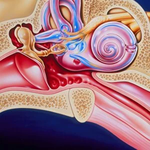
Inner ear hair cells. Confocal light micrograph of V-shaped rows of hair cells (bright arcs) in the organ of Corti. This structure lies in the cochlea of the inner ear, and converts sound vibrations into nerve impulses. The hairs (stereocilia) are embedded in the basilar membrane. When sound waves arrive at this membrane from the middle ear, they cause it to vibrate. This pushes the hair cells against the overlying tectorial membrane (not seen). This triggers nerve impulses, which travel to the brain through the auditory nerve. Magnification unknown
Science Photo Library features Science and Medical images including photos and illustrations
Media ID 6422476
© DAVID BECKER/SCIENCE PHOTO LIBRARY
Auditory Sense Aural Cochlea Cochlear Confocal Light Micrograph Fluorescence Fluorescent Hearing Immunofluorescence Immunofluorescent Organ Of Corti Sense Sensory Stereocilia Stereocilium Nervous System
EDITORS COMMENTS
This print from Science Photo Library showcases the intricate beauty of inner ear sensory hairs, specifically the inner ear hair cells. The image reveals V-shaped rows of these delicate hair cells, forming bright arcs within the organ of Corti. Nestled in the cochlea of the inner ear, this remarkable structure plays a crucial role in converting sound vibrations into nerve impulses. The stereocilia, or hairs, are embedded within the basilar membrane and react to incoming sound waves. As these waves reach the membrane from the middle ear, they set it into motion, causing it to vibrate. Consequently, this movement pushes against an unseen tectorial membrane above and stimulates the hair cells. Through this fascinating mechanism, nerve impulses are triggered and swiftly travel through the auditory nerve towards our brain for interpretation. This process is fundamental to our ability to hear and perceive sounds around us. Immersed in vibrant fluorescence under confocal light microscopy techniques, this micrograph offers a glimpse into one aspect of human anatomy that contributes immensely to our sense of hearing. It highlights not only its scientific significance but also its aesthetic appeal as a work of art captured by Science Photo Library's expertise in visual storytelling.
MADE IN AUSTRALIA
Safe Shipping with 30 Day Money Back Guarantee
FREE PERSONALISATION*
We are proud to offer a range of customisation features including Personalised Captions, Color Filters and Picture Zoom Tools
SECURE PAYMENTS
We happily accept a wide range of payment options so you can pay for the things you need in the way that is most convenient for you
* Options may vary by product and licensing agreement. Zoomed Pictures can be adjusted in the Cart.