Home > Science > Xray
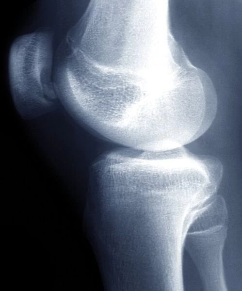
Knee disease, X-ray F006 / 9138
![]()

Wall Art and Photo Gifts from Science Photo Library
Knee disease, X-ray F006 / 9138
Knee disease. Coloured X-ray of the right knee of an 18 year old patient with osteochondritis dissecans (OCD) of the knee. A small piece of bone and cartilage has separated from the rear of the patella (knee cap, upper left). The separated pieces may stay in place or fall into the joint. OCD causes pain and swelling and, if the bone has moved, locking of the joint
Science Photo Library features Science and Medical images including photos and illustrations
Media ID 9252355
© ZEPHYR/SCIENCE PHOTO LIBRARY
Human Body Part Human Leg Illness Knee Cap Patella Radiography Radiological Radiology Scientific Imaging Xray Abnormal Condition Human Knee Unhealthy
FEATURES IN THESE COLLECTIONS
EDITORS COMMENTS
This print showcases the intricate details of a knee disease known as osteochondritis dissecans (OCD). The coloured X-ray image reveals the right knee of an 18-year-old patient, highlighting a small piece of bone and cartilage that has detached from the rear of the patella, or knee cap. Positioned against a striking black background, this close-up shot provides a clear view of the separated fragments, which may either remain in place or fall into the joint. OCD is characterized by pain, swelling, and potential joint locking if the displaced bone has shifted. This condition can cause significant discomfort and hinder mobility for those affected. With its focus on disease and illness within human anatomy, this photograph serves as an invaluable tool for medical professionals seeking to understand and diagnose such conditions accurately. The radiographic quality of this image emphasizes its scientific nature while offering valuable insights into healthcare practices. Radiology plays a crucial role in capturing detailed images like these for diagnostic purposes. By shedding light on abnormal anatomical features through scientific imaging techniques like X-rays, experts can better comprehend various diseases affecting bones and joints. ZEPHYR/SCIENCE PHOTO LIBRARY presents this remarkable visual representation without any commercial intentions but rather with a commitment to advancing medical knowledge surrounding osteochondritis dissecans and related ailments.
MADE IN AUSTRALIA
Safe Shipping with 30 Day Money Back Guarantee
FREE PERSONALISATION*
We are proud to offer a range of customisation features including Personalised Captions, Color Filters and Picture Zoom Tools
SECURE PAYMENTS
We happily accept a wide range of payment options so you can pay for the things you need in the way that is most convenient for you
* Options may vary by product and licensing agreement. Zoomed Pictures can be adjusted in the Cart.

