Home > Popular Themes > Human Body
Lower arm bones and ligaments
![]()

Wall Art and Photo Gifts from Science Photo Library
Lower arm bones and ligaments
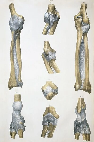
Lower arm bones & ligaments. Historical anatomical artwork of lower arm bones (yellow) and ligaments (pale blue). Ligaments are bands of fibrous tissue that hold bones together at joints. The lower arm contains the ulna and radius bones, and is shown from the front (upper left) and behind (upper right, ulna at left). These join with the humerus (upper arm bone) at the elbow, seen down centre in four views/positions (from top): inner side; outer side; flexed, from behind; extended, from front. The wrist joint is seen from the inner side (ulna at top, lower right) and the outer side (radius at top, lower right). From The Bones and Ligaments of the Human Body (Ed. Jones Quain, London, 1842)
Science Photo Library features Science and Medical images including photos and illustrations
Media ID 6419442
© SHEILA TERRY/SCIENCE PHOTO LIBRARY
1842 Anterior Arthrological Arthrology Bones Book Connective Tissue Dissected Dissection Drawing Elbow Extended Flexed Front Frontal Humerus Joint Joints Jones Quain Ligament Ligaments Lower Radius Skeletal Text Book Ulna Wrist
EDITORS COMMENTS
This historical anatomical artwork showcases the intricate details of lower arm bones and ligaments. The print, dating back to 1842, provides a glimpse into the fascinating world of human anatomy during the 19th century. In this illustration, we can observe the yellow-colored lower arm bones alongside pale blue ligaments that play a crucial role in holding these bones together at joints. Ligaments are depicted as bands of fibrous tissue, emphasizing their connective function. The focus is primarily on the ulna and radius bones found in the lower arm region. The image presents two perspectives: one from the front and another from behind, with an emphasis on the ulna bone on the left side. Moving towards other significant areas, we encounter views of four different positions of the elbow joint along its centerline: inner side, outer side, flexed from behind, and extended from front. These positions highlight various aspects of movement and flexibility within this joint. Additionally, attention is drawn to both sides of the wrist joint – one view showcasing ulna at top (inner side) while another displaying radius at top (outer side). Overall, this remarkable piece offers valuable insights into medical history by presenting detailed illustrations dissecting skeletal structures and providing essential knowledge for students studying medicine or anyone intrigued by human anatomy's intricacies.
MADE IN AUSTRALIA
Safe Shipping with 30 Day Money Back Guarantee
FREE PERSONALISATION*
We are proud to offer a range of customisation features including Personalised Captions, Color Filters and Picture Zoom Tools
SECURE PAYMENTS
We happily accept a wide range of payment options so you can pay for the things you need in the way that is most convenient for you
* Options may vary by product and licensing agreement. Zoomed Pictures can be adjusted in the Cart.

