Home > Popular Themes > Human Body
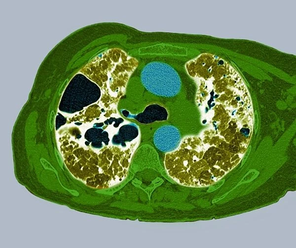
Lung fibrosis, CT scan
![]()

Wall Art and Photo Gifts from Science Photo Library
Lung fibrosis, CT scan
Lung fibrosis. Image 2 of 3. Coloured computed tomography (CT) scan through the chest of a patient with lung (pulmonary) fibrosis. This is the middle of the lung with the aortic branches (blue), non-lung tissue (green), lung tissue (yellow/brown) and air-filled bullae (black). Lung fibrosis is a condition whereby a lung infection causes inflammation of the tissue around the alveoli (air sacs), producing scar tissue. This scarring leads to breathlessness due to stiffnesss of the lungs. Damaged areas of the alveoli also fill with air, forming bullae or blebs. If they burst, the air released causes pneumothorax (collapsed lung), which can be life-threatening. See M160/059 and 061 for the rest of the sequence
Science Photo Library features Science and Medical images including photos and illustrations
Media ID 6414148
© DU CANE MEDICAL IMAGING LTD/SCIENCE PHOTO LIBRARY
Alveoli Alveolitis Alveolus Blebs Blister Blisters Bullae Chest Collapsed Computed Tomography Ct Scan False Colour Infection Inflamed Inflammation Lung Lungs Middle Section Pathological Pathology Pneumothorax Pulmonary Respiration Respiratory System Sacs Scanner Scar Tissue Scarring Stiffness Abnormal Condition False Coloured Unhealthy
EDITORS COMMENTS
This print showcases a coloured computed tomography (CT) scan of a patient's chest, specifically focusing on lung fibrosis. The image reveals the intricate details of the middle section of the lungs, highlighting various elements such as aortic branches in blue, non-lung tissue in green, lung tissue in yellow/brown, and air-filled bullae in black. Lung fibrosis is an unfortunate condition that occurs when inflammation caused by a lung infection affects the tissue surrounding the alveoli or air sacs. This inflammation leads to the formation of scar tissue, resulting in stiffness and breathlessness for individuals affected by this condition. Additionally, damaged areas within the alveoli fill with air and form bullae or blebs. If these structures burst, it can lead to pneumothorax or collapsed lung which poses life-threatening risks. The image not only provides valuable insight into this pathological state but also highlights its potential consequences. By understanding these complexities through CT scans like this one, medical professionals gain crucial information necessary for diagnosis and treatment planning. Science Photo Library has expertly captured this visually striking representation of pulmonary fibrosis using false colours to enhance clarity and comprehension. It serves as a reminder of both the fragility and resilience of our respiratory system while emphasizing ongoing research efforts towards finding curative solutions for interstitial lung diseases like fibrosis.
MADE IN AUSTRALIA
Safe Shipping with 30 Day Money Back Guarantee
FREE PERSONALISATION*
We are proud to offer a range of customisation features including Personalised Captions, Color Filters and Picture Zoom Tools
SECURE PAYMENTS
We happily accept a wide range of payment options so you can pay for the things you need in the way that is most convenient for you
* Options may vary by product and licensing agreement. Zoomed Pictures can be adjusted in the Cart.

