Home > Popular Themes > Human Body
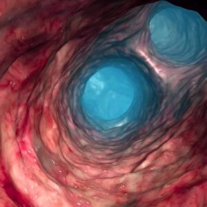
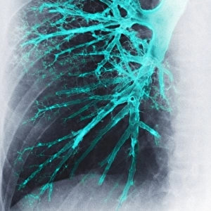
Lung, X-ray

![]()
Wall Art and Photo Gifts from Science Photo Library
Lung, X-ray
Lung. Coloured bronchography (X-ray) of a healthy human lung. A contrast medium has been added to show the network of airways (orange) in the right lung. The trachea (wind pipe) enters the lungs and splits into the two bronchi (one seen, upper right). The bronchi then further divide into bronchioles, which continue to divide and decrease in diameter. The bronchioles eventually end in groups of tiny air sacs known as alveoli (not seen). Alveoli are the site of gaseous exchange between the lungs and blood; oxygen enters the blood and carbon dioxide leaves
Science Photo Library features Science and Medical images including photos and illustrations
Media ID 6449981
© CNRI/SCIENCE PHOTO LIBRARY
Airway Airways Branches Branching Breathing Bronchi Bronchiole Bronchioles Bronchus Chest Contrast Medium False Colour Gas Exchange Gaseous Exchange Lung Net Work Pulmonary Radio Opaque Radiography Respiration Respiratory Ribs System Technique Thoracic Thorax Trachea X Ray Machine False Coloured
MADE IN AUSTRALIA
Safe Shipping with 30 Day Money Back Guarantee
FREE PERSONALISATION*
We are proud to offer a range of customisation features including Personalised Captions, Color Filters and Picture Zoom Tools
SECURE PAYMENTS
We happily accept a wide range of payment options so you can pay for the things you need in the way that is most convenient for you
* Options may vary by product and licensing agreement. Zoomed Pictures can be adjusted in the Cart.