Home > Popular Themes > Human Body
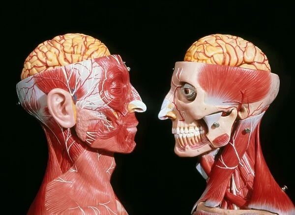
Models showing the cerebrum, facial & neck muscles
![]()

Wall Art and Photo Gifts from Science Photo Library
Models showing the cerebrum, facial & neck muscles
Models showing the location of the facial and neck muscles. The top part of the skulls has also been removed to reveal the cerebrum together with the blood vessels which provide its blood supply. The temporalis, a fan-shaped muscle responsible for the movements of the mandible, is seen on the model at right. The sternocleidomastoid muscle is seen in the neck; it has a cylindrical shape and runs obliquely from the posterior lower part of the cranium to the anterior part of the collar bone. The trapezium extends over the back of the neck and controls the position of the shoulder blade during movement. The parotid gland is seen close to the ear on the model at left
Science Photo Library features Science and Medical images including photos and illustrations
Media ID 6452205
© GEOFF TOMPKINSON/SCIENCE PHOTO LIBRARY
EDITORS COMMENTS
This print from Science Photo Library showcases the intricate details of the human body's facial and neck muscles. The models on display provide a comprehensive view of these vital structures, offering an enlightening insight into their location and function. The focal point of this image lies in the removal of the top part of the skulls, which unveils the cerebrum alongside its intricate network of blood vessels responsible for nourishing this essential organ. This unique perspective highlights both the complexity and beauty that lie within our brains. Moving towards the right model, we encounter the temporalis muscle, resembling a fan shape. This powerful muscle plays a crucial role in mandible movement, allowing us to chew and speak with precision. Shifting our attention to the neck area, we are introduced to two prominent muscles: sternocleidomastoid and trapezium. The cylindrical-shaped sternocleidomastoid runs obliquely from behind lower cranium to collar bone anteriorly, contributing significantly to head rotation and flexion movements. Meanwhile, trapezium extends over the back of our necks while controlling shoulder blade positioning during various bodily motions. Lastly, near one model's ear is an intriguing sight -the parotid gland. Positioned close by but often overlooked due to its small size; it serves as a critical salivary gland involved in digestion processes. Through this remarkable photograph print by Science Photo Library, viewers gain valuable insights into human anatomy—unveiling not only how our muscles work
MADE IN AUSTRALIA
Safe Shipping with 30 Day Money Back Guarantee
FREE PERSONALISATION*
We are proud to offer a range of customisation features including Personalised Captions, Color Filters and Picture Zoom Tools
SECURE PAYMENTS
We happily accept a wide range of payment options so you can pay for the things you need in the way that is most convenient for you
* Options may vary by product and licensing agreement. Zoomed Pictures can be adjusted in the Cart.

