Home > Science > SEM
Nerve fibres
![]()

Wall Art and Photo Gifts from Science Photo Library
Nerve fibres
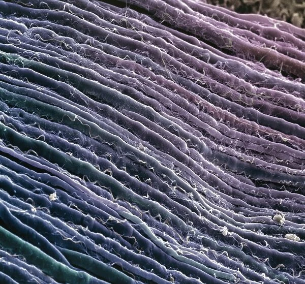
Nerve fibres. Coloured scanning electron micrograph (SEM) of parallel myelinated nerve fibres in the spinal cord. Each fibre consists of a nerve cell axon, the output process of a nerve cell, surrounded by a fatty insulating layer known as a myelin sheath. Myelin sheaths increase the transmission speed of the electrical nerve signals. Nerves in the body serve to collect, interpret and relay information. The fibres pass the information on to other parts of the body, such as the muscles and organs. The spinal cord, housed within the vertebrae of the backbone, links the brain to the rest of the body. Magnification: x650 at 6x7cm size
Science Photo Library features Science and Medical images including photos and illustrations
Media ID 6421772
© STEVE GSCHMEISSNER/SCIENCE PHOTO LIBRARY
Axon Axons Connective Tissue Fibre Fibres Groove Grooves Histological Histology Line Lines Myelin Myelinated Nerve Nerve Fibre Nerves Nervous Parallel Spinal Cord System White Matter
FEATURES IN THESE COLLECTIONS
EDITORS COMMENTS
This print showcases the intricate network of nerve fibres found in the spinal cord. The coloured scanning electron micrograph (SEM) beautifully captures the parallel arrangement of myelinated nerve fibres, each consisting of a nerve cell axon enveloped by a protective myelin sheath. The presence of these myelin sheaths is crucial as they significantly enhance the speed at which electrical signals are transmitted along the nerves. Acting as information highways, these fibres play a vital role in collecting, interpreting, and relaying sensory and motor information throughout our body. As we delve deeper into understanding the human nervous system, this image serves as a visual reminder of its complexity and importance. These fibres act as messengers, passing on valuable information to various parts of our body including muscles and organs. Situated within the vertebrae of our backbone, the spinal cord acts as an essential link between our brain and other bodily systems. It serves as a central hub for transmitting signals that allow us to move, feel sensations, and carry out countless other functions necessary for daily life. With its magnification at x650 in 6x7cm size, this SEM image offers us an up-close glimpse into one aspect of our incredible anatomy – reminding us just how remarkable and interconnected our bodies truly are.
MADE IN AUSTRALIA
Safe Shipping with 30 Day Money Back Guarantee
FREE PERSONALISATION*
We are proud to offer a range of customisation features including Personalised Captions, Color Filters and Picture Zoom Tools
SECURE PAYMENTS
We happily accept a wide range of payment options so you can pay for the things you need in the way that is most convenient for you
* Options may vary by product and licensing agreement. Zoomed Pictures can be adjusted in the Cart.


