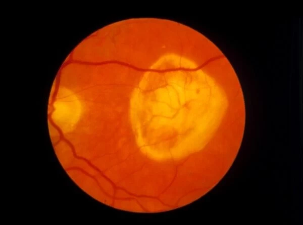
Ophthalmoscopy of disciform macula degeneration
![]()

Wall Art and Photo Gifts from Science Photo Library
Ophthalmoscopy of disciform macula degeneration
Macula degeneration. Ophthalmoscope view of the retina of a patients eye, showing disciform macula degeneration. At centre right is a large disc-shaped discoloured area covering the macula, the part of the retina responsible for acuity of vision. Retinal cells responsible for detecting images of light are destroyed and there may be breakthrough to the choroid layer beneath. At right is the yellow optic disc where blood vessels and the optic nerve enter the eye. Macula degeneration can occur due to trauma, but most commonly is a progressive disorder in the elderly. The effect is loss of central vision. Laser treat- ment may be successful if diagnosed early
Science Photo Library features Science and Medical images including photos and illustrations
Media ID 6422703
© PAUL PARKER/SCIENCE PHOTO LIBRARY
Eye Disease Ophthalmic Ophthalmoscopy Retina Condition Disorder Health Care
EDITORS COMMENTS
This print from Science Photo Library showcases the ophthalmoscopic view of a patient's eye affected by disciform macula degeneration. The image reveals a striking disc-shaped discolored area at the center-right, enveloping the macula - the vital part of the retina responsible for sharp vision. Unfortunately, this condition leads to destruction of retinal cells that detect light images and may even penetrate into the choroid layer beneath. On the right side, we can observe the yellow optic disc where blood vessels and the optic nerve enter. Disciform macula degeneration can be caused by trauma but is predominantly seen as a progressive disorder in older individuals. Its detrimental consequence manifests as central vision loss, significantly impacting daily life activities. However, there is hope for those diagnosed early with laser treatment proving successful in some cases. Science Photo Library presents this remarkable photograph to shed light on this disease and its effects on our visual health. It serves as an educational tool for medical professionals and researchers studying ophthalmic conditions such as macular degeneration or fundoscopy.
MADE IN AUSTRALIA
Safe Shipping with 30 Day Money Back Guarantee
FREE PERSONALISATION*
We are proud to offer a range of customisation features including Personalised Captions, Color Filters and Picture Zoom Tools
SECURE PAYMENTS
We happily accept a wide range of payment options so you can pay for the things you need in the way that is most convenient for you
* Options may vary by product and licensing agreement. Zoomed Pictures can be adjusted in the Cart.


