Home > Science > SEM
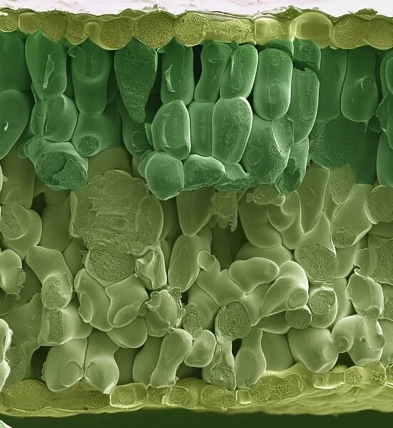
SEM of spinach leaf
![]()

Wall Art and Photo Gifts from Science Photo Library
SEM of spinach leaf
Spinach leaf. Coloured scanning electron micrograph (SEM) of a fractured leaf of the spinach plant, Spinacia oleracea. At top and bottom frame are a single layer of cells (light green) which form the epidermis of the leaf. The leaf interior contains palisade mesophyll (dark green) & spongy mesophyll (light green). The tightly packed palisade cells contain chloroplasts, sites of photosynthesis. The spongy mesophyll cells have with fewer chloroplasts; they are more involved with gas exchange. Magnification: x219 at 6x7cm size
Science Photo Library features Science and Medical images including photos and illustrations
Media ID 9194285
© POWER AND SYRED/SCIENCE PHOTO LIBRARY
Epidermis Mesophyll Palisade Mesophyll Plants Spinach Spinacia Oleracea Spongy Mesophyll
EDITORS COMMENTS
This print showcases the intricate beauty of a spinach leaf at a microscopic level. The image, captured using a scanning electron microscope (SEM), reveals the complex structure and functionality of this common plant. At first glance, we are drawn to the vibrant colors that dominate the frame. The top and bottom edges display a single layer of light green cells forming the epidermis, which acts as a protective outer covering for the leaf. Moving inward, we encounter two distinct regions: palisade mesophyll and spongy mesophyll. The dark green palisade mesophyll cells immediately catch our attention due to their tightly packed arrangement. These specialized cells contain an abundance of chloroplasts, responsible for conducting photosynthesis – nature's remarkable process by which plants convert sunlight into energy. Their strategic positioning near the upper surface allows them to efficiently capture sunlight for maximum productivity. Contrasting with their densely packed counterparts, the light green spongy mesophyll cells exhibit fewer chloroplasts but play an essential role in gas exchange within the leaf. This region facilitates efficient diffusion of carbon dioxide and oxygen between stomata on the leaf's surface and surrounding tissues. With its magnification set at 219 times its original size, this SEM image provides us with an extraordinary glimpse into one small aspect of nature's grand design – reminding us that even seemingly ordinary objects like spinach leaves hold incredible complexity when observed up close.
MADE IN AUSTRALIA
Safe Shipping with 30 Day Money Back Guarantee
FREE PERSONALISATION*
We are proud to offer a range of customisation features including Personalised Captions, Color Filters and Picture Zoom Tools
SECURE PAYMENTS
We happily accept a wide range of payment options so you can pay for the things you need in the way that is most convenient for you
* Options may vary by product and licensing agreement. Zoomed Pictures can be adjusted in the Cart.


