Home > Science > SEM
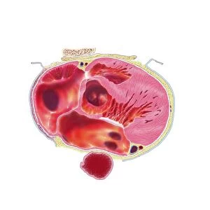
Seminiferous tubule of testis

![]()
Wall Art and Photo Gifts from Science Photo Library
Seminiferous tubule of testis
Seminiferous tubule. Coloured scanning electron micrograph (SEM) of a sectioned seminiferous tubule, the site of sperm production in the human testis. At the centre of the tubule, sperm cells are forming whose hair-like tails are seen. Each testis is packed with seminiferous tubules. The tubule is lined with epithelium containing two types of cell (blue/yellow): Sertoli cells which nourish developing sperm, and spermatogenic cells which produce the sperm. The sperm are released into the cavity of the tubule to migrate to the epididymis, where they mature. Around the seminiferous tubule are Leydig cells (pink) which produce testosterone. Magnification: unknown
Science Photo Library features Science and Medical images including photos and illustrations
Media ID 6450435
© STEVE GSCHMEISSNER/SCIENCE PHOTO LIBRARY
Re Production Reproductive System Seminiferous Tubule Site Sperm Spermatogenesis Spermiogenesis Testis
FEATURES IN THESE COLLECTIONS
EDITORS COMMENTS
This print from Science Photo Library offers a mesmerizing glimpse into the intricate world of human reproduction. The image showcases a sectioned seminiferous tubule, which serves as the primary site for sperm production within the testis. Packed with countless tubules, each testis is a bustling hub of activity. The seminiferous tubule featured in this photograph is lined with epithelium containing two distinct types of cells – Sertoli cells and spermatogenic cells. The Sertoli cells play a vital role in nourishing and supporting the developing sperm, while the spermatogenic cells are responsible for producing these tiny, life-giving entities. At the center of the tubule, one can observe an awe-inspiring sight: sperm cells in various stages of formation. These miniature marvels possess hair-like tails that will eventually enable them to swim towards their destination – the epididymis – where they will undergo further maturation. Surrounding this dynamic scene are Leydig cells, depicted in a striking pink hue. These specialized cells produce testosterone, an essential hormone involved in male reproductive function. Through its meticulous detail and vibrant colors, this scanning electron micrograph invites us to appreciate both the complexity and beauty inherent within our own bodies' reproductive systems. It serves as a testament to science's ability to capture moments that would otherwise remain hidden from our naked eyes - truly showcasing how artistry can intertwine with scientific exploration.
MADE IN AUSTRALIA
Safe Shipping with 30 Day Money Back Guarantee
FREE PERSONALISATION*
We are proud to offer a range of customisation features including Personalised Captions, Color Filters and Picture Zoom Tools
SECURE PAYMENTS
We happily accept a wide range of payment options so you can pay for the things you need in the way that is most convenient for you
* Options may vary by product and licensing agreement. Zoomed Pictures can be adjusted in the Cart.