Home > Science > SEM
Small intestine villi, SEM

![]()
Wall Art and Photo Gifts from Science Photo Library
Small intestine villi, SEM
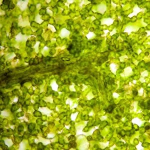
Small intestine villi. Coloured scanning electron micrograph (SEM) of villi (folds) on the lining of the small intestine. Villi greatly increase the intestinal surface area for absorbing nutrients from food. The epithelium (outer layer, folded) of each villus contains enterocyte cells, which are involved in nutrient absorption, and goblet cells, which secrete mucus onto the intestinal surface. The inner villus tissue (not seen) contains blood vessels which transport digestive products to a nearby vein. Magnification: x27 at 6x7cm size
Science Photo Library features Science and Medical images including photos and illustrations
Media ID 6450175
© SUSUMU NISHINAGA/SCIENCE PHOTO LIBRARY
Absorption Absorptive Alimentary Canal Digestion Digestive System Lining Mucosa Mucosal Nutrient Projection Projections Secretory Site Small Intestine Surface Area Tissue Tract Villi Villus
FEATURES IN THESE COLLECTIONS
EDITORS COMMENTS
This print showcases the intricate beauty of small intestine villi, as captured by a coloured scanning electron micrograph (SEM). The folds and projections on the lining of the small intestine form these remarkable structures known as villi. These tiny finger-like protrusions play a crucial role in our digestive system. The epithelium, or outer layer, of each villus is folded and consists of enterocyte cells responsible for absorbing nutrients from our food. Additionally, goblet cells are present which secrete mucus onto the intestinal surface to aid in digestion. The inner tissue of the villus contains blood vessels that efficiently transport digested products to nearby veins. What makes these villi truly remarkable is their ability to greatly increase the surface area of the small intestine for nutrient absorption. This adaptation ensures that our bodies can effectively extract essential nutrients from ingested food. This SEM image magnified at x27 reveals stunning details that highlight both the complexity and functionality of this vital part of our anatomy. It serves as a reminder of how intricately designed our bodies are to carry out normal healthy digestion. Science Photo Library has once again provided us with an awe-inspiring glimpse into nature's wonders through this incredible photograph print.
MADE IN AUSTRALIA
Safe Shipping with 30 Day Money Back Guarantee
FREE PERSONALISATION*
We are proud to offer a range of customisation features including Personalised Captions, Color Filters and Picture Zoom Tools
SECURE PAYMENTS
We happily accept a wide range of payment options so you can pay for the things you need in the way that is most convenient for you
* Options may vary by product and licensing agreement. Zoomed Pictures can be adjusted in the Cart.