Home > Popular Themes > Human Body
Wuchereria bancrofti parasite
![]()

Wall Art and Photo Gifts from Science Photo Library
Wuchereria bancrofti parasite
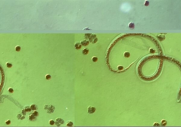
Wuchereria bancrofti. Light micrograph of the microfilaria larval stage of the parasitic worm Wuchereria bancrofti, which causes filariasis in humans. W. bancrofti larvae are also found in several mosquito hosts. They enter the human body as the mosquitoes feed, and migrate to the lymphatic system, where they mature. The adult female produces thousands of microfilariae, which enter the circulation and are ingested by feeding mosquitoes. Adult worms can block the lymph vessels, causing thickening and enlargement of infected tissues. Other symptoms include fever and swelling of the lymph nodes. Differential interference contrast. Magnification unknown
Science Photo Library features Science and Medical images including photos and illustrations
Media ID 6470167
© SINCLAIR STAMMERS/SCIENCE PHOTO LIBRARY
Contrast Diagnosis Diagnostic Differential Interference Infection Infectious Larva Lymphatic Nematoda Nematode Parasite Parasitic Round Worm Vector Borne Worm Light Micrograph
EDITORS COMMENTS
This print showcases the intricate world of parasites, specifically the Wuchereria bancrofti parasite. The image reveals a light micrograph of the microfilaria larval stage of this parasitic worm, which is responsible for causing filariasis in humans. Wuchereria bancrofti larvae can be found residing within various mosquito hosts. As these mosquitoes feed on human blood, they introduce the larvae into our bodies. From there, these tiny creatures embark on a journey through our lymphatic system to mature and reproduce. The adult female Wuchereria bancrofti produces an astonishing number of microfilariae that enter our circulation and are subsequently ingested by feeding mosquitoes, perpetuating their life cycle. However, this parasitic relationship comes at a cost to us as hosts. Adult worms have the ability to obstruct lymph vessels, leading to thickening and enlargement of infected tissues. Alongside this distressing consequence, symptoms such as fever and swollen lymph nodes may also manifest. Through differential interference contrast microscopy techniques employed in capturing this image, we gain insight into the fascinating yet unsettling nature of vector-borne infections caused by nematodes like Wuchereria bancrofti. This remarkable photograph serves as both a diagnostic tool for medical professionals studying filariasis and an awe-inspiring reminder of the intricate complexities present within nature's realm.
MADE IN AUSTRALIA
Safe Shipping with 30 Day Money Back Guarantee
FREE PERSONALISATION*
We are proud to offer a range of customisation features including Personalised Captions, Color Filters and Picture Zoom Tools
SECURE PAYMENTS
We happily accept a wide range of payment options so you can pay for the things you need in the way that is most convenient for you
* Options may vary by product and licensing agreement. Zoomed Pictures can be adjusted in the Cart.

