Home > Animals > Mammals > Vespertilionidae > Intermedius
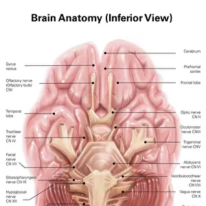
Anatomy of human brain stem and cranial nerves

![]()
Wall Art and Photo Gifts from Stocktrek
Anatomy of human brain stem and cranial nerves
Stocktrek Images specializes in Astronomy, Dinosaurs, Medical, Military Forces, Ocean Life, & Sci-Fi
Media ID 13009835
© Alan Gesek/Stocktrek Images
Anatomy Biology Biomedical Illustrations Brain Brain Stem Central Nervous System Cerebellum Cerebrum Communication Cranial Nerves Diagram Directly Below Facial Nerves Frontal Lobe Healthcare Human Anatomy Human Body Parts Human Brain Intelligence Medical Medicine Medulla Nerve Nervous System Neuroanatomy Neurology Neuroscience Optic Nerves Peripheral Nervous System Physiology Sensory System Spinal Nerves Temporal Lobe Text Western Script Abducens Nerve Hypoglossal Nerve Oculomotor Nerve Trigeminal Nerve Trochlear Nerve Vagus Nerve
FEATURES IN THESE COLLECTIONS
> Animals
> Mammals
> Muridae
> Intermedius
> Animals
> Mammals
> Vespertilionidae
> Intermedius
> Arts
> Street art graffiti
> Digital art
> Digital paintings
EDITORS COMMENTS
This vibrant and detailed print captures the intricate beauty of the human brain stem and cranial nerves. Against a clean white background, this vertical artwork showcases biomedical illustrations that are digitally generated in stunning color. With no people present, the focus is solely on the complex network of nerves that play a crucial role in our communication and overall well-being. From the trigeminal nerve to the trochlear nerve, from facial nerves to vagus nerve, each component is meticulously depicted with precision. The vestibulocochlear nerves, abducens nerve, oculomotor nerve, and more are all visually represented alongside their corresponding labels in Western script. This illustration not only highlights neuroanatomy but also emphasizes its significance within healthcare and medicine. It serves as a reminder of how essential understanding our own physiology is for diagnosing and treating various conditions related to our central nervous system. The image encompasses key structures such as the cerebellum, cerebrum, medulla, temporal lobe, frontal lobe, infundibulum, gyrus rectus - all integral parts contributing to intelligence and sensory systems. Created by Alan Gesek exclusively for Stocktrek Images (not mentioning commercial use), this diagram provides an invaluable resource for those studying neuroscience or simply fascinated by human anatomy. Whether displayed in medical facilities or educational institutions alike- it serves as a visual testament to our incredible complexity as sentient beings.
MADE IN AUSTRALIA
Safe Shipping with 30 Day Money Back Guarantee
FREE PERSONALISATION*
We are proud to offer a range of customisation features including Personalised Captions, Color Filters and Picture Zoom Tools
SECURE PAYMENTS
We happily accept a wide range of payment options so you can pay for the things you need in the way that is most convenient for you
* Options may vary by product and licensing agreement. Zoomed Pictures can be adjusted in the Cart.