Home > Arts > Street art graffiti > Digital art > Digital paintings
Ewings sarcoma locations on the skeleton

![]()
Wall Art and Photo Gifts from Stocktrek
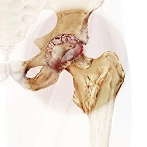
Ewings sarcoma locations on the skeleton
Ewings sarcoma locations on the skeleton and detail of tumor on head of femur
Stocktrek Images specializes in Astronomy, Dinosaurs, Medical, Military Forces, Ocean Life, & Sci-Fi
Media ID 13008607
© TriFocal Communications/Stocktrek Images
Anatomy Appendicular Skeleton Biology Biomedical Illustrations Bone Cancer Cancer Cells Clavicle Cortex Costae Spuriae Costae Verae Cranium Damaged Detail Disease Disorder False Ribs Femur Fibula Floating Ribs Healthcare Human Anatomy Human Bones Human Skeleton Human Tissue Humerus Illness Intercostal Space Lumbar Vertebrae Malignant Medical Medicine Oncology Osteology Pathology Pelvis Physiology Pubic Bones Rib Cage Skeletal System Skeleton Sternum Text Thoracic Cage Tibia True Ribs Tumor Ulna Unhealthy Vertebrochondral Ribs Vertebrosternal Ribs Western Script Periosteum
FEATURES IN THESE COLLECTIONS
> Arts
> Street art graffiti
> Digital art
> Digital paintings
EDITORS COMMENTS
This digitally generated image showcases the locations of Ewings sarcoma on the human skeleton, with a detailed focus on a tumor found on the head of the femur. The illustration, presented in vibrant colors against a white background, provides valuable insight into this rare form of cancer that primarily affects children and young adults. The skeletal system is meticulously depicted, highlighting key anatomical structures such as the periosteum and cortex. Various bones are labeled including the humerus, femur, pelvis, tibia, fibula, ulna, sternum, clavicle, and cranium. Additionally, different types of ribs are identified - floating ribs (costae spuriae), false ribs (vertebrochondral ribs), and true ribs (costae verae) - along with their placement within the intercostal spaces forming the thoracic cage. The image serves as an educational tool for medical professionals and students studying oncology or pathology. It emphasizes how Ewings sarcoma can manifest in different regions throughout the appendicular skeleton and lumbar vertebrae. By providing a visual representation of this devastating disease at both macroscopic and microscopic levels through damaged bone tissue invaded by malignant cancer cells; it underscores its impact on overall health while raising awareness about early detection methods crucial to successful treatment outcomes. TriFocal Communications has expertly crafted this biomedical illustration to contribute to our understanding of Ewings sarcoma's pathological effects within our bodies' intricate skeletal framework.
MADE IN AUSTRALIA
Safe Shipping with 30 Day Money Back Guarantee
FREE PERSONALISATION*
We are proud to offer a range of customisation features including Personalised Captions, Color Filters and Picture Zoom Tools
SECURE PAYMENTS
We happily accept a wide range of payment options so you can pay for the things you need in the way that is most convenient for you
* Options may vary by product and licensing agreement. Zoomed Pictures can be adjusted in the Cart.